0226
Storing phase information in the longitudinal direction: Experimental verification of double diffusion encoding with stimulated echoes applied to closed pores1Medical Physics in Radiology, German Cancer Research Center (DKFZ), Heidelberg, Germany, 2Faculty of Physics and Astronomy, Heidelberg University, Heidelberg, Germany, 3Institute of Radiology, University Hospital Erlangen, Friedrich-Alexander-Universität Erlangen-Nürnberg (FAU), Erlangen, Germany
Synopsis
When applying double diffusion encoding (DDE) to arbitrarily shaped closed pores, non-vanishing imaginary parts in the diffusion signal arise, which allow determining the average shape of the pores in the considered volume element. Key limitations in such experiments are the available gradient strength and reaching the diffusion long-time limit restricted by T2 decay. When incorporating stimulated echoes into DDE-sequences, the slower T1 relaxation can be exploited to reach the long-time limit in larger pores demanding lower gradient strengths. We present experimental verification that phase information can be stored in longitudinal magnetization direction thus preserving complex signals under application of stimulated echoes.
Introduction
Diffusion pore imaging enables imaging of the average shape of arbitrary closed pores for investigation of the microstructure of biological tissue and porous materials. To measure the Fourier transform $$$\tilde{\rho}(\boldsymbol{q})$$$ of the pore space function $$$\rho(\boldsymbol{x})$$$, non-conventional gradient profiles are used: Either a long-narrow gradient profile can be applied to measure $$$\tilde{\rho}(\boldsymbol{q})$$$ directly1-5. Alternatively, q-space imaging providing the magnitude $$$|\tilde{\rho}(\boldsymbol{q})|$$$ can be combined with double diffusion encoded (DDE) measurements from which the phase of $$$\tilde{\rho}(\boldsymbol{q})$$$ can be extracted6-8. Both methods involve measurements of complex signals containing the shape information of non-point-symmetric pores, which is lost in classical diffusion encoding schemes.
While the long-narrow approach has a rather inflexible sequence design, the second method uses only short gradient pulses and allows incorporating stimulated echoes in-between gradients to reach the diffusion long-time limit by exploiting the longer T1-relaxation time compared to T2. This might enable diffusion pore imaging in larger pores with lower demand on gradient strength, which reduces quadratically with an increase in pore diameter.
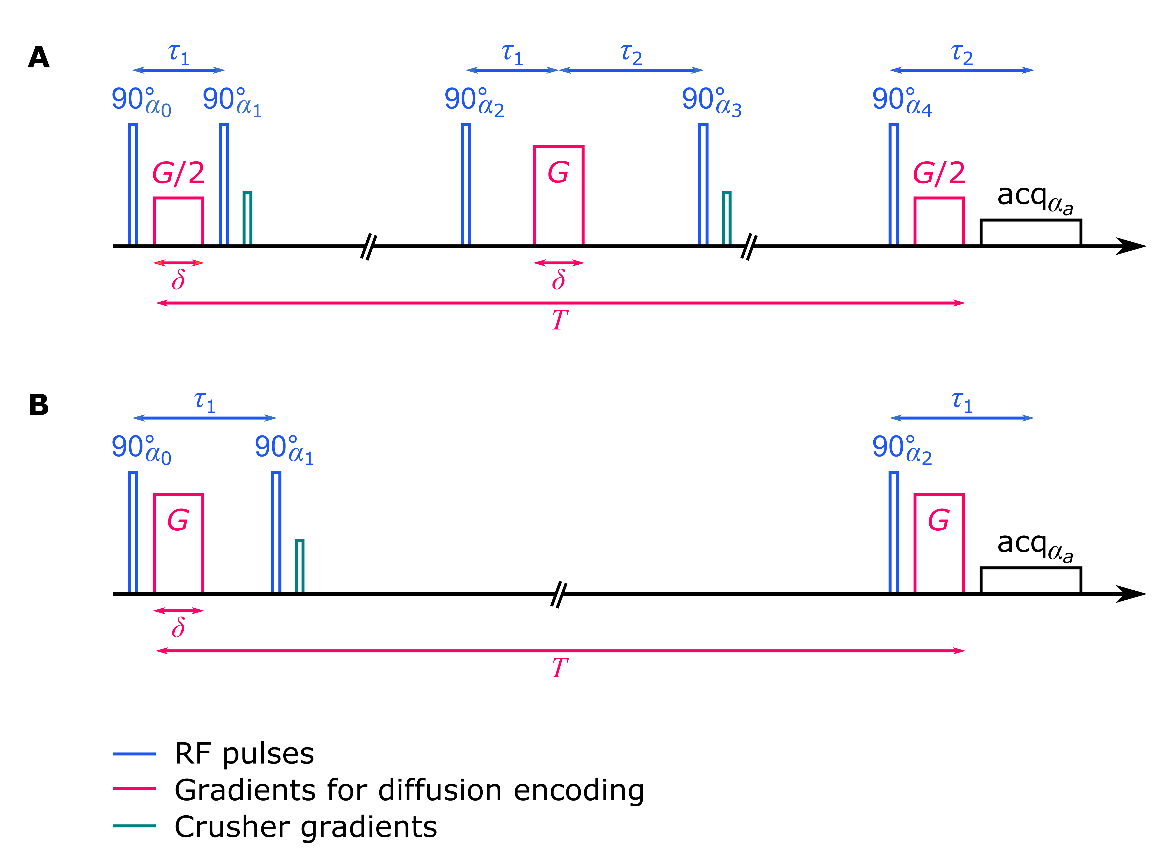
So far, only real signals have been acquired with stimulated echoes. In this study, we present a first experimental verification for storing complex signals in longitudinal magnetization direction for the case of a double diffusion encoded stimulated echo sequence involving five RF pulses (Fig. 1A).
Theory
When using stimulated echoes, the DDE-signal consists of four terms9,10: the desired signal term, that also arises for a spin echo sequence, plus three undesired terms modulated with the Fourier transform of the pore center of mass distribution $$$\tilde{P}_{x_{\text{cm}}}(\boldsymbol{q})$$$,$$S_{\text{DDE}}(\boldsymbol{q})=\frac{1}{4}\,e^{i(\alpha_0-\alpha_1-\alpha_2+\alpha_3+\alpha_4)}\,\tilde{\rho}^*(\boldsymbol{q}/2)^2\tilde{\rho}(\boldsymbol{q})\,+\,\frac{1}{4}\,e^{i(\alpha_0-\alpha_1+\alpha_2-\alpha_3+\alpha_4)}\,\tilde{P}^*_{x_{\text{cm}}}(2\boldsymbol{q})\,\tilde{\rho}^*(\boldsymbol{q}/2)^2\tilde{\rho}^*(\boldsymbol{q})\,+\\\frac{1}{4}\,e^{i(-\alpha_0+\alpha_1-\alpha_2+\alpha_3+\alpha_4)}\,\tilde{P}_{x_{\text{cm}}}(\boldsymbol{q})\,|\tilde{\rho}(\boldsymbol{q}/2)|^2\tilde{\rho}(\boldsymbol{q})\,+\,\frac{1}{4}\,e^{i(-\alpha_0+\alpha_1+\alpha_2-\alpha_3+\alpha_4)}\,\tilde{P}^*_{x_{\text{cm}}}(\boldsymbol{q})\,|\tilde{\rho}(\boldsymbol{q}/2)|^2\tilde{\rho}^*(\boldsymbol{q})\text{,}$$ with RF-pulse phases $$$\alpha_i$$$ (Fig. 1), q-space vector $$$\boldsymbol{q}=\gamma\boldsymbol{G}\delta$$$, gyromagnetic ratio $$$\gamma$$$, gradient pulse amplitude $$$\boldsymbol{G}$$$ and duration $$$\delta$$$, and the asterisk indicating complex conjugates. In most samples, $$$\tilde{P}_{x_{\text{cm}}}(\boldsymbol{q})$$$ should decay to zero already at small q values $$$|\boldsymbol{q}|$$$ and hence no deviation of the stimulated echo signal from the spin echo signal should be observed10 apart from the reduced amplitude and a phase depending on the choice of RF-pulse phases, yielding $$$S_{\text{DDE}}(\boldsymbol{q})=\frac{1}{4}\,e^{i(\alpha_0-\alpha_1-\alpha_2+\alpha_3+\alpha_4)}\,\tilde{\rho}^*(\boldsymbol{q}/2)^2\tilde{\rho}(\boldsymbol{q})$$$. Similarly, the stimulated echo signal for q-space imaging reduces from two terms to $$$S_{\text{QSI}}(\boldsymbol{q})=\frac{1}{2}\,e^{i(-\alpha_0+\alpha_1+\alpha_2)}\,|\tilde{\rho}(\boldsymbol{q})|^2$$$.
Methods
A phantom with 77 pores of equilateral triangular shape (5.75 mm edge length) was used, for which $$$\tilde{P}_{x_{\text{cm}}}(\boldsymbol{q})$$$ decays to zero quickly. Hyperpolarized Xe-129 gas generated by spin exchange optical pumping was used as the diffusing medium inside the pores. The stimulated echo sequences (Fig. 1) were implemented on a clinical 1.5 T MR-scanner (Magnetom Symphony, Siemens).
For q-space imaging with three RF pulses five echoes occur, but for the 5-pulse DDE sequence 68 echo pathways arise. To measure only the desired stimulated echo, RF spoiling and gradient spoiling were used. RF-pulse phases were chosen following Zur et al.11: $$$\alpha_0$$$ =117, $$$\alpha_1$$$=234, $$$\alpha_2$$$=108, $$$\alpha_3$$$=99, $$$\alpha_4$$$=207. The crusher gradients were implemented in the z-storage periods of the desired stimulated echo.
$$$S_\text{QSI}(q)$$$ and
$$$S_\text{DDE}(q)$$$ were recorded along the vertical gradient
direction.
$$$\tilde{\rho}(q)$$$ was calculated by recursively estimating the
phase of
$$$\tilde{\rho}(q)$$$ from
$$$S_\text{DDE}(q)$$$ and using
$$$|\tilde{\rho}(q)|=\sqrt{S_\text{QSI}(q)}$$$ for the magnitude7. An inverse Fourier transform then yields
$$$\rho(x)$$$. This was repeated for 37 radial spokes in q space followed by an inverse Radon transform to
reconstruct a two-dimensional image. For comparison, simulations of the
diffusion process were conducted using a matrix approach to solve the
Bloch-Torrey equations2,12.
Experimental results
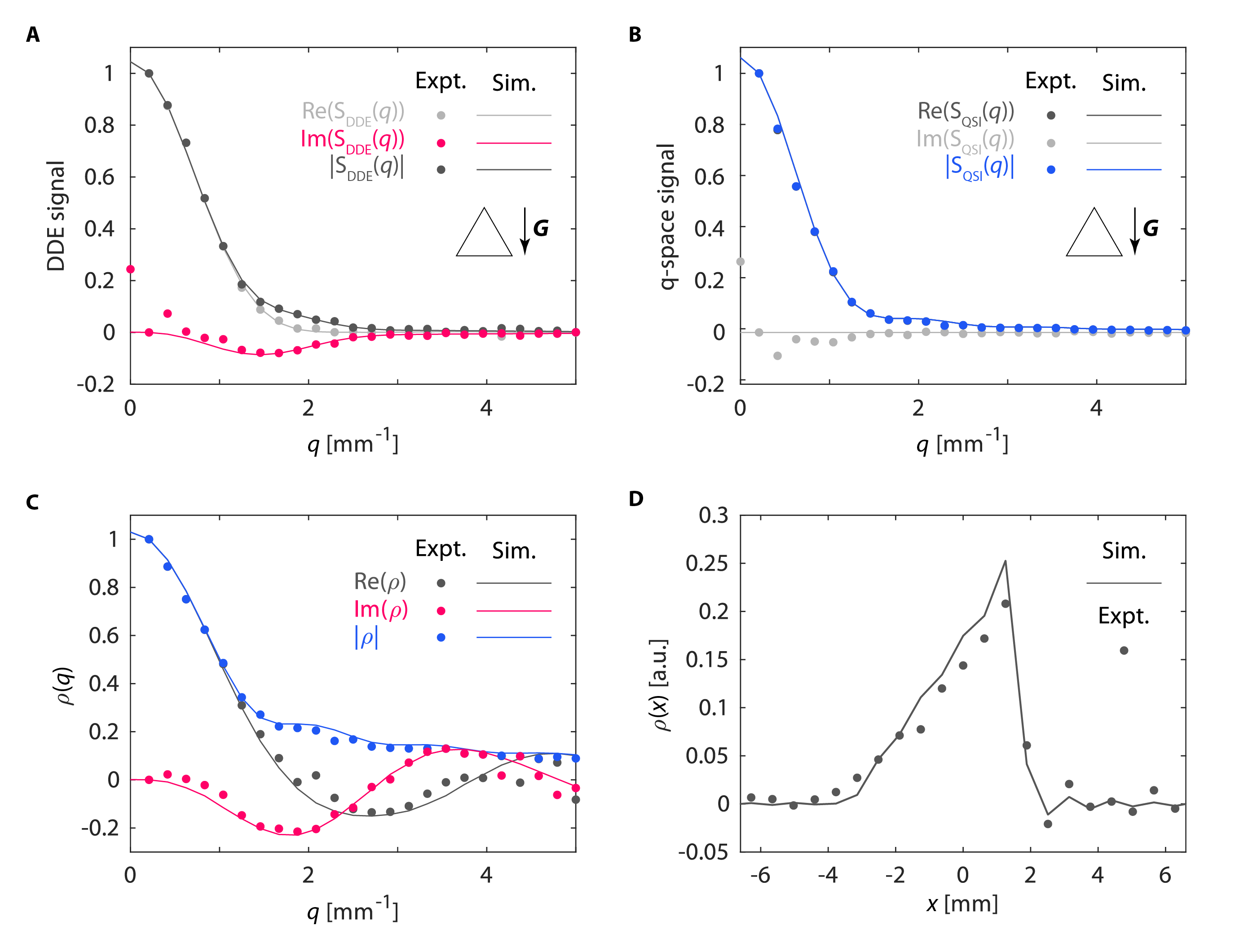
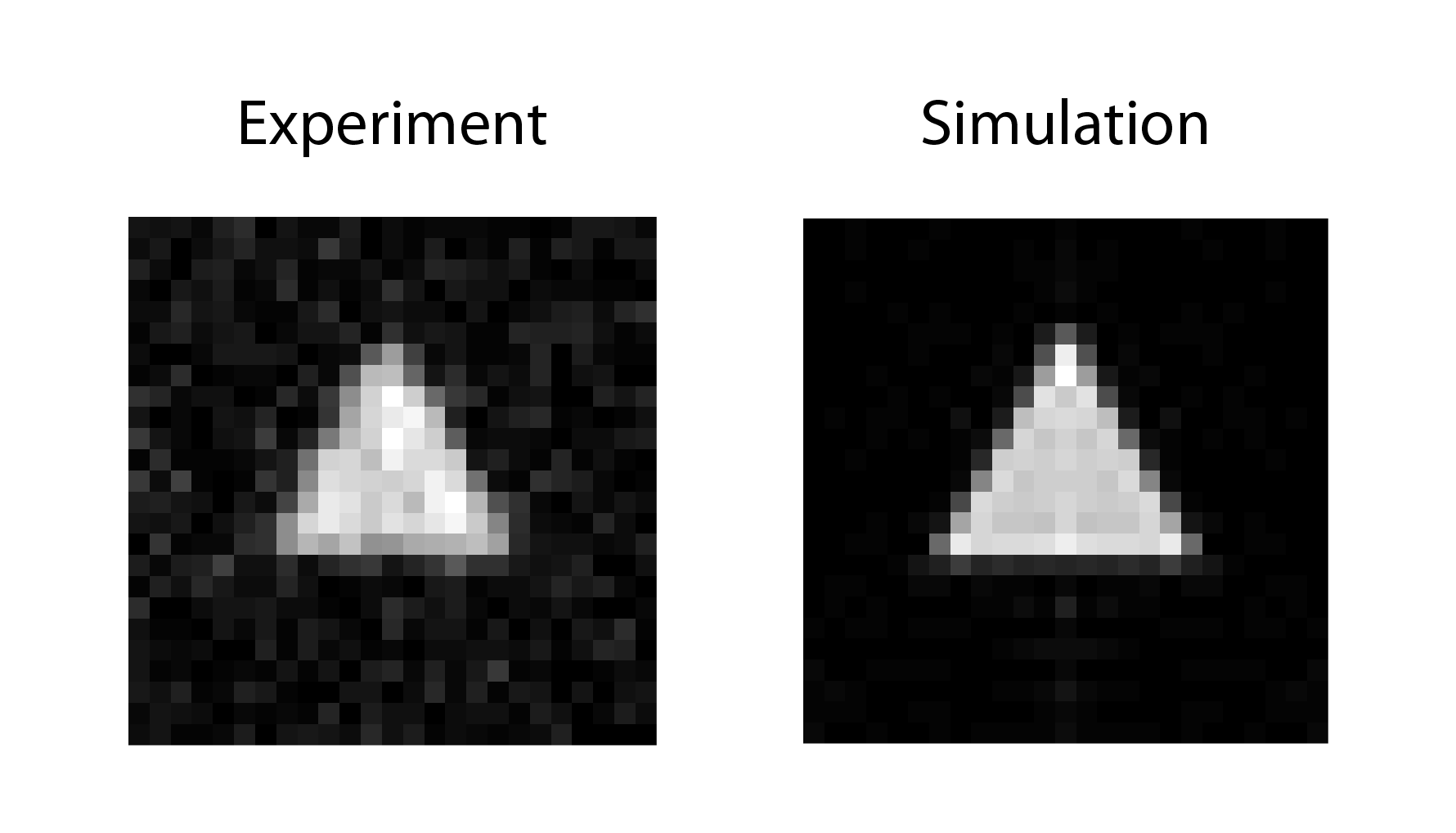
Real and imaginary parts of the measured signals $$$S_\text{DDE}(q)$$$ and $$$S_\text{QSI}(q)$$$ are shown in Fig. 2A-B. Due to the non-point-symmetry of the triangle, non-vanishing imaginary parts in the DDE signal arise (Fig. 2A, magenta), which allows recursively estimating the phase of $$$\tilde{\rho}(q)$$$ (Fig. 2C). The inverse Fourier transform of $$$\tilde{\rho}(q)$$$ corresponds to the projection of the triangular shape onto the gradient direction (Fig. 2D). The stimulated echo signals agree well with the simulations, except for small deviations in the imaginary parts at low q values. These are also responsible for blurring the pore image, but the triangular shape is still clearly discernible (Fig. 3).
Discussion
These initial experiments demonstrate that the phase information of complex signals is preserved in stimulated echoes. Simulations for different absolute phantom positions suggest that the $$$\tilde{P}_{x_{\text{cm}}}(\boldsymbol{q})$$$-terms might have a small contribution to the signal. If confirmed, phase cycling can be used to remove them10. Otherwise further adjustments of the measurement sequences are required to yield decent phase stability.Conclusion
Applicability of stimulated echoes is in principle of interest to all fields where signal phases arise. With regard to diffusion pore imaging, the demands on gradient strength might be met on whole-body scanners for imaging of muscle cells, with much larger diameters than most cells (~50 µm), using phosphocreatine as the tracer. In muscle tissue, T2 is quite short unlike T1, so that stimulated echoes may be used as a tool to reach the diffusion long-time limit.
Acknowledgements
Financial support by the DFG (grant no. KU 3362/1-1 and LA 2804/6-1) is gratefully acknowledged.References
1) Laun F.B., Kuder T.A. et al., Determination of the defining boundary in nuclear magnetic resonance
diffusion experiments, Phys. Rev. Lett. 107, 048102 (2011).
2) Laun F.B., Kuder T.A. et al., NMR-based diffusion pore imaging, Phys. Rev. E 86, 021906 (2012).
3) Kuder T.A., Bachert
P. et al., Diffusion pore imaging by hyperpolarized xenon-129 nuclear magnetic
resonance, Phys. Rev. Lett. 111, 028101 (2013).
4) Hertel S., Hunter
M. et al., Magnetic resonance pore imaging, a tool for porous media research,
Phys. Rev. E 87, 030802 (2013).
5) Hertel S.A., Wang X. et al., Magnetic-resonance pore imaging of nonsymmetric microscopic pore shapes,
Phys. Rev. E 92, 012808 (2015).
6) Shemesh N., Westin
C.F. et al., Magnetic resonance imaging by synergistic diffusion-diffraction
patterns, Phys. Rev. Lett. 108, 058103 (2012).
7) Kuder T.A. and Laun F.B., NMR-based diffusion pore imaging by
double wave vector measurements, Magn. Reson. Med. 70, 836-841 (2013).
8) Demberg K., Laun
F.B. et al., Nuclear magnetic resonance diffusion pore imaging: Experimental phase
detection by double diffusion encoding, Phys. Rev. E 95, 022404 (2017).
9) Khrapitchev A.A.
and Callaghan P.T., Double PGSE NMR with
Stimulated Echoes: Phase Cycles for the Selection of Desired Encoding, J.
Magn. Reson. 152, 259-268 (2001).
10) Demberg K., Laun F.B. et al., Validity extension of stimulated echoes to imaginary signals arising
for double diffusion encoding of closed pores, Proc. Intl. Soc. Mag. Reson.
Med. 26, 1103 (2018).
11) Zur Y., Wood M.L. et al., Spoiling of transverse magnetization in steady-state sequences,
Magn. Reson. Med. 21, 251-263 (1991).
12)
Grebenkov D.S., NMR survey of
reflected Brownian motion, Rev. Mod. Phys. 79, 1077 (2007).
Figures