0225
SPatiotemporal ENcoding (SPEN) at ultra-high fields: Applications to high resolution (<100 µm isotropic) in vivo mouse brain DTI1Chemical and Biological Physics, Weizmann Institute of Science, Rehovot, Israel, 2Champalimaud Centre for the Unknown, Lisbon, Portugal
Synopsis
SPEN can be a valuable alternative to Spin Echo EPI, especially at very high fields where sensitivity is maximized but magnetic field susceptibility artifacts become important. This work presents a new Paravision® 6 software package capable of acquiring and processing SPEN data, incorporating single/multishot acquisitions, motion correction, image zooming and multiple contrast possibilities such as CEST, diffusion or multi-echo acquisition. The power of this method is exemplified with in vivo 2D and 3D diffusion tensor imaging (DTI) studies on whole and zoomed mouse brain regions, acquired at 15.2T utilizing a cryoprobe and reaching isotropic resolution of 75 µm.
INTRODUCTION
Echo Planar Imaging (EPI) remains a key component in a wide range of applications requiring fast MRI, including functional imaging and Diffusion Weighted / Diffusion Tensor Imaging (DWI / DTI). EPI, however, faces known challenges when targeting heterogenous tissues, when operating at high magnetic fields, or in other instances where field inhomogeneities become important. A number of features make Spatiotemporal Encoding (SPEN) a robust alternative to deal with these challenges1–3:
1) SPEN can be implemented in a fully T2* refocused manner where inhomogeneities are compensated for throughout the acquisition.
2) Its bandwidth along the blipped dimension –the more artifact-prone in EPI– is defined at the excitation stage by a chirp pulse that can be set at arbitrary values.
3) SPEN directly records its images in spatial space along the low-bandwidth dimension, and, since each signal yields a direct low-resolution image, referenceless motion correction between shots becomes facile and data free from motion artifacts in multishot and/or interleaved acquisitions can be obtained4–6.
4) SPEN’s use of a chirp pulse applied in conjunction with an encoding gradient induces a spatial selectivity, that permits zooming without folding along the low-bandwidth SPEN dimension.
Some of these advantages are illustrated for high field in vivo mouse brain imaging in Figure 1. This work introduces an acquisition and processing package that exploits these advantages within the context of preclinical scanning in a Bruker Paravision® 6 environment to perform SPEN-based in vivo 3D DTI of mice brain at 15.2T.
METHODS
The SPEN and SE EPI pulse sequences used in this study, both with multislice and 3D encoding, are shown in Figure 2. The SPEN package (running on custom-written code in Paravision® 6 environment), is available for download (see below) and includes the possibility of super-resolution processing, motion correction, and multishot acquisitions6 as well as numerous contrast possibilities such as CEST, diffusion7, multi-echo acquisition, fat suppression, etc. The resulting SPEN images are compared to Spin Echo EPI results with double sampling8 (state-of-the-art in current scanners).RESULTS & DISCUSSION
Figure 3 compare in vivo 2D multislice DTI results of an in vivo mouse brain collected with SPEN, Spin Echo and SE EPI with double sampling (dS). The SPEN and SE EPI images reach a spatial resolution of 75x75x500 µm. The quality and SNR of all image are excellent. The SPEN images show reduced inhomogeneities artifacts, e.g., near the ear canals, compared to SE EPI. This is despite that the bandwidths used for the multishot EPI and SPEN experiments were very similar (10.4 and 11.3 kHz respectively). This favorable performance originates from the fully T2*-refocused nature of SPEN acquisitions.
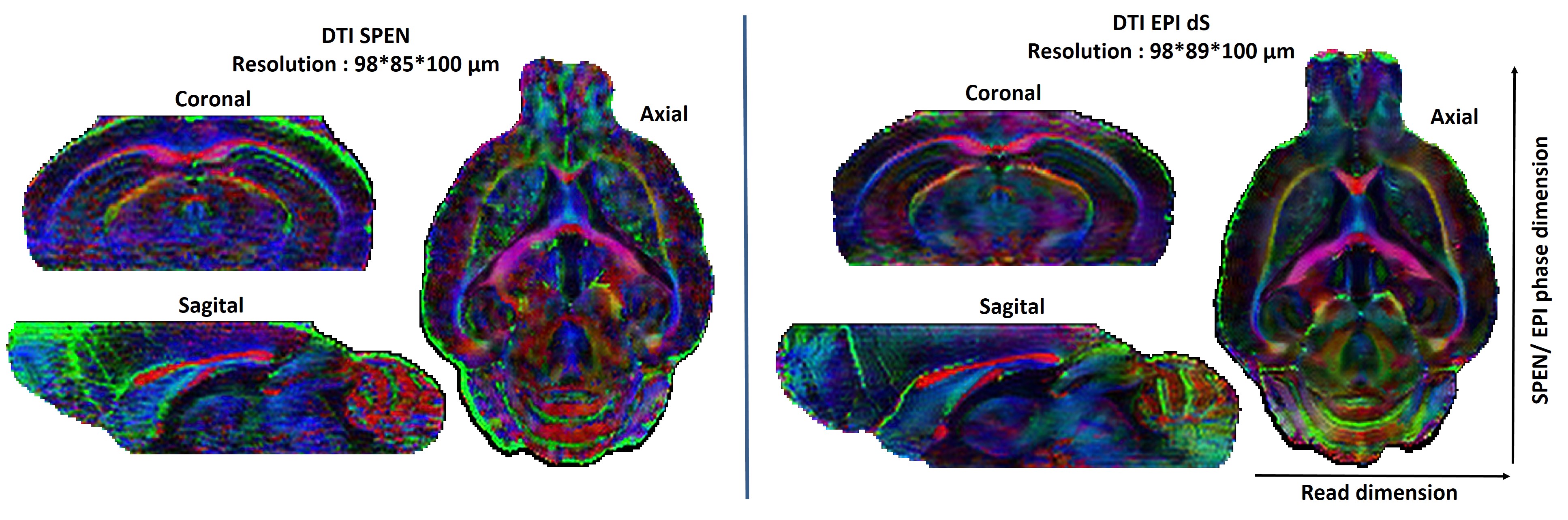
Figure 4 shows color-coded main diffusion direction extracted from in vivo 3D DTI data of a mouse brain acquired in two hours with a resolution of ≈ 100 µm isotropic. The SPEN and the SE EPI images exhibit similar morphological details. SPEN images have a slightly lower SNR than EPI due to their longer echo times and higher effective b-values induced by the SPEN imaging gradient; still, the SPEN images also show reduced inhomogeneities artifacts –particularly in the posterior cerebellum region.
As mentioned, one of SPEN’s advantages rests in its ability to zoom on specific area of the brain and thus increase the resolution achievable in a given acquisition time. Figure 5 demonstrates this potential with four color-coded main diffusion direction slices extracted from an in vivo 3D DTI experiment, focused solely on the cerebellum region of a mouse brain. The resolution is 75 µm isotropic, and allows one to reach a level of detail which has not been reported in vivo before9.
CONCLUSION
The SPEN acquisition and processing software package available in https://www.weizmann.ac.il/chemphys/Frydman_group/software paves the way for preclinical microDTI, functional MRI, and real-time acquisitions in very high magnetic fields with high resolution and robustness.Acknowledgements
We are grateful to Dr. Tangi Roussel (Weizmann/CEA), Dr. Yas Tesiram (Univ Queensland), Dr. Sonia Goncalves (Champalimaud) and Dr. Sascha Koehler (Bruker BioSpin) for their help with the programming. The authors also acknowledge the support from the Israel Science Foundation (grants 2508/17 and 965/18), the Kimmel Institute for Magnetic Resonance (Weizmann Institute) and the generosity of the Perlman Family Foundation.References
1. Ben-Eliezer, N. & Frydman, L. Spatiotemporal encoding as a robust basis for fast three-dimensional in vivo MRI. NMR Biomed. 24, 1191–1201 (2011).
2. Schmidt, R. & Frydman, L. New spatiotemporal approaches for fully refocused, multislice ultrafast 2D MRI. Magn. Reson. Med. 71, 711–722 (2014).
3. Solomon, E., Liberman, G., Nissan, N. & Frydman, L. Robust diffusion tensor imaging by spatiotemporal encoding: Principles and in vivo demonstrations. Magn. Reson. Med. 77, 1124–1133 (2017).
4. Seginer, A., Schmidt, R., Leftin, A., Solomon, E. & Frydman, L. Referenceless reconstruction of spatiotemporally encoded imaging data: Principles and applications to real-time MRI. Magn. Reson. Med. 72, 1687–1695 (2014).
5. Schmidt, R., Seginer, A. & Frydman, L. Interleaved multishot imaging by spatiotemporal encoding: A fast, self-referenced method for high-definition diffusion and functional MRI. Magn. Reson. Med. 75, 1935–1948 (2016).
6. Bao, Q., Liberman, G., Solomon, E., Lustig, M. & Fydman, L. Diffusion-weighted in vivo imaging with =100 μm resolution: principles and applications to ADC mapping of pregnant mice. Proc. Intl. Soc. Mag. Res. Med. 1021 (2018). at <ismrm 2018 1021>
7. Solomon, E., Shemesh, N. & Frydman, L. Diffusion weighted MRI by spatiotemporal encoding: Analytical description and in vivo validations. J. Magn. Reson. 232, 76–86 (2013).
8. Yang, Q. X., Posse, S., Bihan, D. L. E. & Smith, M. B. Double-sampled echo-planar imaging at 3 tesla. J. Magn. Reson. - Ser. B 113, 145–150 (1996).
9. Wu, D. et al. In vivo high-resolution diffusion tensor imaging of the mouse brain. Neuroimage 83, 18–26 (2013).
Figures