0221
B-0 shimming of the liver using a local array of shim coils in the presence of respiratory motion at 7T1Center for Image Sciences, UMC Utrecht, Utrecht, Netherlands, 2MR Shim GmbH, Reutlingen, Germany
Synopsis
Inhomogeneity of the magnetic field (B0) in the human body, for instance caused by physiological motion (e.g. breathing), can lead to artifacts. Static second order magnetic field shimming even in the absence of breathing cannot provide a uniform magnetic field in large organs such as the liver, particularly at ultra-high field throughout the breathing cycle. We show that in conjunction with static second order shimming, local arrays of shim coils can substantially improve the magnetic field uniformity in the liver at different breathing states.
Introduction
Variations of the static magnetic field (B0) in the human body can lead to serious frequency offsets during MR and magnetic resonance spectroscopy. If not accounted for, this can be reflected in the form of artifacts, peak shifts, line broadening, and signal loss.1 These variations are partially caused by physiological motion like breathing, and other temporal B0 field fluctuations in the MR system. Higher magnetic field strengths, such as 7T, are more sensitive to the effects of large dynamic B0 field variations. Applying image based B0 shimming, while monitoring breathing and/or respiratory triggering, helps in further reducing the B0 field variations, however, usually at the cost of an extended scan time. In this study, we show that variations in B0 can be reduced at different respiratory states where the static field is kept homogeneous (i.e. B0 shimmed). In conjunction with static second order shimming, with the addition of a local array of shim coils that can be steered externally to adjust the residual magnetic field inhomogeneities, the field homogeneity can be improved throughout the liver.2Methods
4 healthy subjects were scanned at a 7T MR scanner (Philips, Cleveland, USA) after giving informed consent. Eight transceiver fractionated dipole antennas with 16 additional receive loops (MR Coils BV, Zaltbommel, The Netherlands) interfaced to 8 parallel 2kW channels were positioned symmetrically around the abdomen.3 To maximize and homogenize the B1+ field in the liver region, RF phase shimming was performed. Four dual echo B0 maps (GE, 240×304×18mm3 FOV, 2×2×2mm3 voxel size, FA=5$$$^\circ$$$, TR=10ms) during free breathing and three during breath-hold in expiration and inhalation states (GE, 386×410×180mm3 FOV, 6×6×6mm3 voxel size, FA=4$$$^\circ$$$, TR=6ms) were acquired to assess the potential effect of the shim coil array on B0 shimming on the liver (MR Shim GmbH, Reutlingen, Germany). The local shim coil array used for this simulation consisted of 16 circular loops of each 5cm in diameter. The diameters were optimized using a brute-force algorithm between the ranges of 1cm to 10cm. The objective function was defined as the standard deviation of the residual frequency shifts on a sample in vivo dataset. The coils were arranged in two rows of 8 around the torso, (8 on top and 8 at the bottom) (Figure 1). The coil centers of the two rows were 11cm apart. Firstly, shimmed B0 fields were simulated using only static second order shimming (using the free-breathing field maps as reference maps). Secondly, in addition to the static second order shims, the local shim array was used to shim each of the different states of the breathing cycle (using the reference maps acquired from each state separately). Another two B0 simulations were performed: 1) second order for each breathing state, and 2) second order + local shim array for each breathing state. Slice-based analysis was performed to calculate B0 distributions and standard deviations in the three orthogonal directions.Results
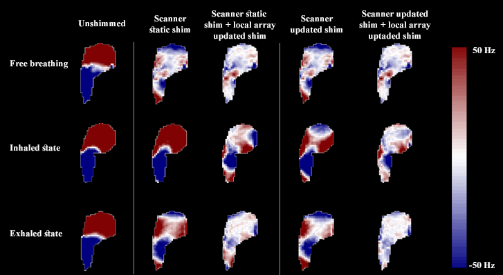
Figure 2 lists that inhaled state B0 maps shows the most improvement when a local shim array is used, because of the large inhomogeneity in the unshimmed B0 maps. Notably, exhaled state and free breathing B0 shims with the local shim coil array show an almost similar improvement of approximately 20%, as compared to the scanner shim. However, when driving both the second order and local shim arrays with per breathing stage optimized shim currents, the exhaled state gives most improvement. Furthermore, the improvement using the lower resolution B0 maps as reference maps is similar to when using higher resolution maps. Figure 3 shows clearly that the local array of shim coils outperforms the scanner B0 shim. Figure 4 shows an example of a slice in the middle of the liver of the B0 field of subject 3 for the four shimming comparisons.Discussion
As expected, there is a large inhomogeneity found in the fully inhaled state B0 maps, due to the amount of air in the lungs. However, the local array of shim coils in conjunction with the second order shimming of the MRI system, can improve the B0 field homogeneity compared to solely using the scanner shim. The inhaled and exhaled state of the breath-hold are the worst case scenarios.Conclusion
This study shows that B0 variations in the liver at 7T can be reduced by an additional 20% after static B0 shimming, even in the presence of respiratory motion by using a local array of statically driven shim coils. For optimal B0 homogeneity, shim updating during different phases of respiratory motion is necessary.Acknowledgements
European H2020-FETOPEN: NICIReferences
1. Truong Trong-Kha, et al. Effects of static and radiofrequency magnetic field inhomogeneity in ultra-high field magnetic resonance imaging. Magnetic Resonance Imaging. 2006;24:103-112.
2. Juchem, Christoph, et al. Dynamic shimming of the human brain at 7 T. Concepts in Magnetic Resonance Part B: Magnetic Resonance Engineering 2010;37(3): 116-128.
3. Raaijmakers A.J.E., et al. The fractionated dipole antenna: A new antenna for body imaging at 7 Tesla. Magnetic Resonance in Medicine. 2016;75:1366–1374.
Figures


Average improvement when using the local array shim for each breathing state compared with the unshimmed and scanner static/updated shimmed B0 maps in percentages during the three breathing states in the coronal, sagittal, and transverse planes of the liver. Static shim used the shim values calculated by the free breathing and updated shim used shim values calculated by all the states separately.
* Only static shimming was performed on the 2mm isotropic B0 map, as no inhaled and exhaled B0 map to perform updated shim by the scanner, were acquired.