0219
Dynamic Multi-Coil Technique (DYNAMITE) MRI on Human Brain1Departments of Biomedical Engineering and Radiology, Columbia University, New York, NY, United States, 2Department of Radiology, Center for Magnetic Resonance Research (CMRR), University of Minnesota, Minneapolis, MN, United States, 3Department of Radiology and Biomedical Imaging, Magnetic Resonance Resesarch Center (MRRC), Yale University, New Haven, CT, United States
Synopsis
To date, spatial encoding for human MRI is based on linear X, Y and Z field gradients generated by dedicated X, Y and Z gradient coils. We recently introduced the Dynamic Multi-Coil Technique (DYNAMITE) for B0 magnetic field modeling and demonstrated DYNAMITE MRI in a miniaturized setup. In this study we report the first realization of multi-slice DYNAMITE MRI of the in vivo human brain in which all gradient fields are purely DYNAMITE-based. The obtained image fidelity is comparable to MRI with conventional gradient coils, paving the way for full-fledged human DYNAMITE MRI systems.
INTRODUCTION
Spatial encoding for magnetic resonance imaging (MRI) is traditionally based on magnetic field gradients generated by dedicated X, Y and Z wire patterns. The DYNAmic Multi-coIl TEchnique (DYNAMITE)1,2 provides a new and generalized method for the synthesis of advanced magnetic field distributions with generic (i.e. non-orthogonal) basis fields from a set of localized coils. DYNAMITE MRI in which all encoding fields are purely multi-coil-generated has been shown in miniaturized setups for radial MRI3, Cartesian MRI and EPI4, and non-linear encoding5 as well as for the concurrent generation of both MRI and B0 shim fields.4 Here we report the first realization of DYNAMITE MRI of the in vivo human brainMETHODS
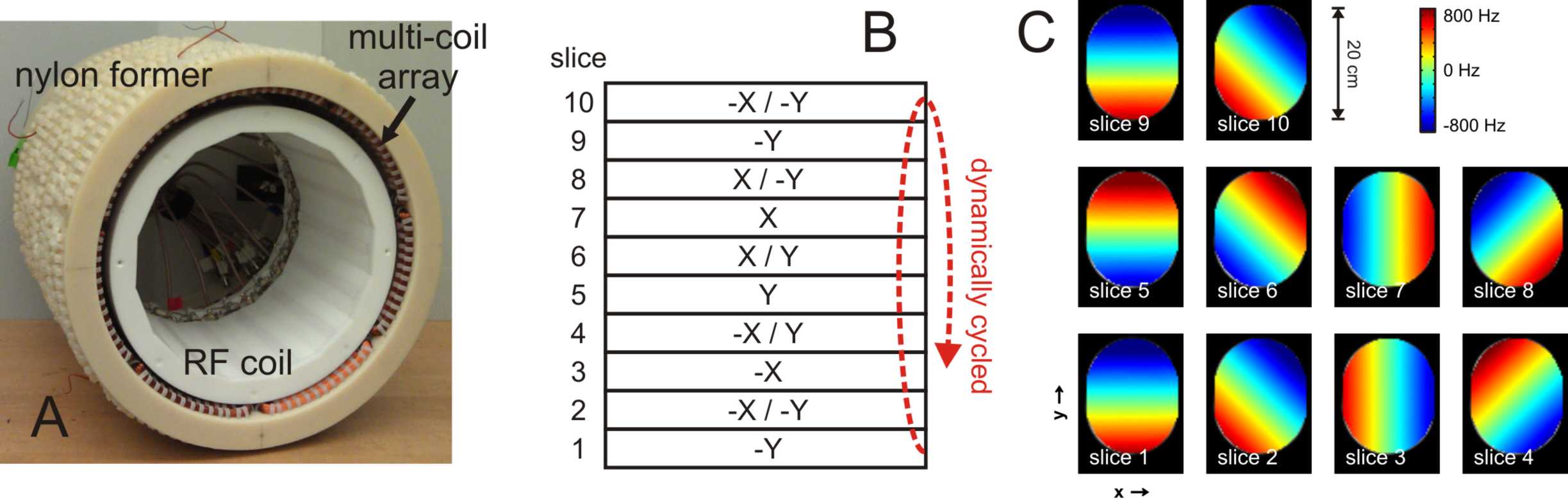
All experiments were performed at Yale University on a 4T Magnex 94 cm magnet interfaced to a Bruker spectrometer. DYNAMITE magnetic fields were generated with a 28 coil matrix distributed as 4 rows of 7 coils on a cylindrical former (Ø 344 mm, spanning 359 mm along the z direction). Each DC coil was composed of two nested rectangular elements of 100 windings each with center dimensions 144 x 80 mm and 118 x 54 mm run in series and interfaced to a ±5 A constant current amplifier (Resonance Research Inc, Billerica, MA). DYNAMITE field modeling and synthesis of the event files for rotating gradient shapes (10 events, figure 1), gradient-echo MRI (18432 events, figure 2) and spin-echo MRI (22528 events) was achieved with customized B0DETOX software.6 The amplitudes and timings of the 28 channels were set by a multi-channel gradient waveform controller, developed in-house.7 RF pulse transmission and reception was achieved with a 16-channel stripline RF coil.8 The calibration of the multi-coil B0 field modeling system and basic functionality testing was done in a head-sized oil phantom (rounded in-plane: 160 x 200 mm, height 20 cm). DYNAMITE MR images were acquired in 5 healthy volunteers with gradient-echo (TR/TE = 1000/14 ms) and spin-echo (TR = 2500/50 ms) sequences as 128 x 128 matrices over 256 x 256 mm (16 slices over 3 mm) and compared to conventional MRI at identical parameters employing the scanner's built-in XYZ gradient coils.RESULTS
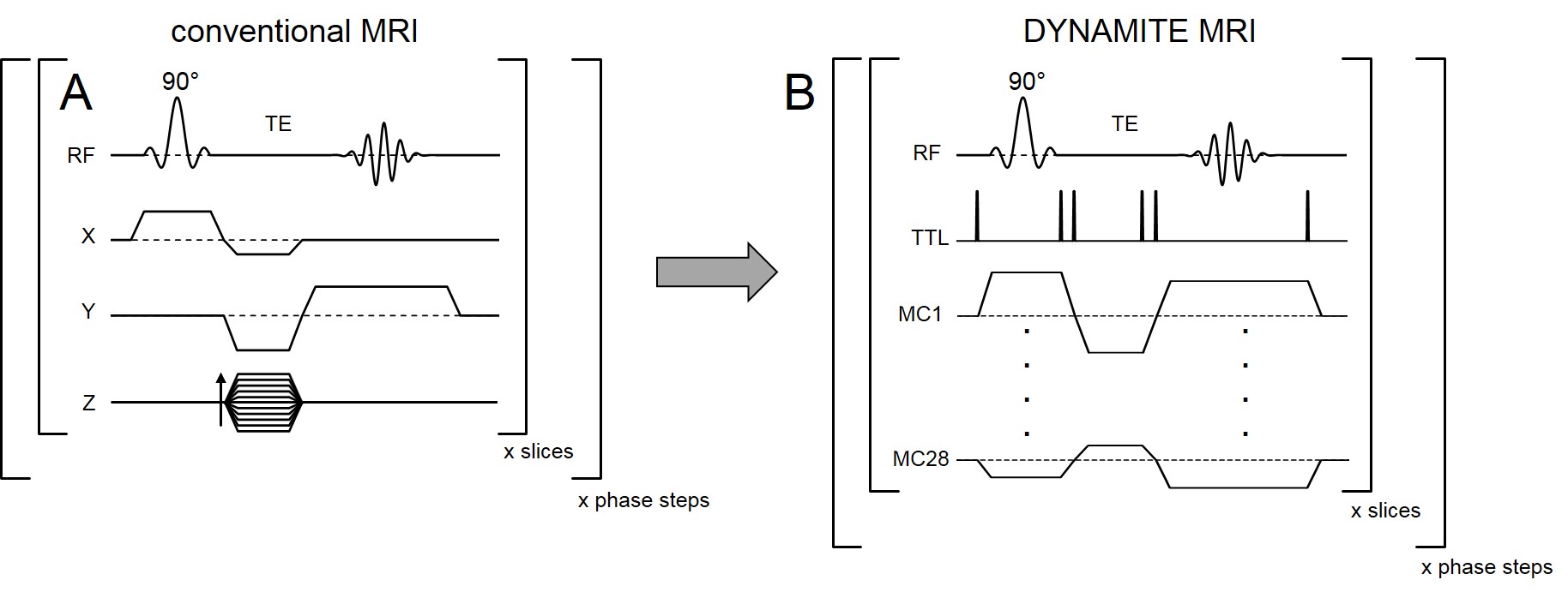
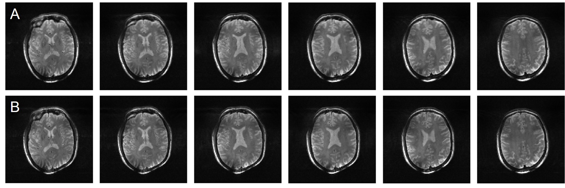
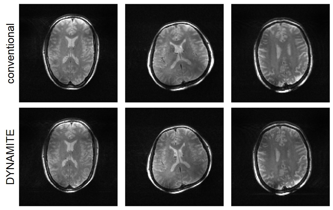
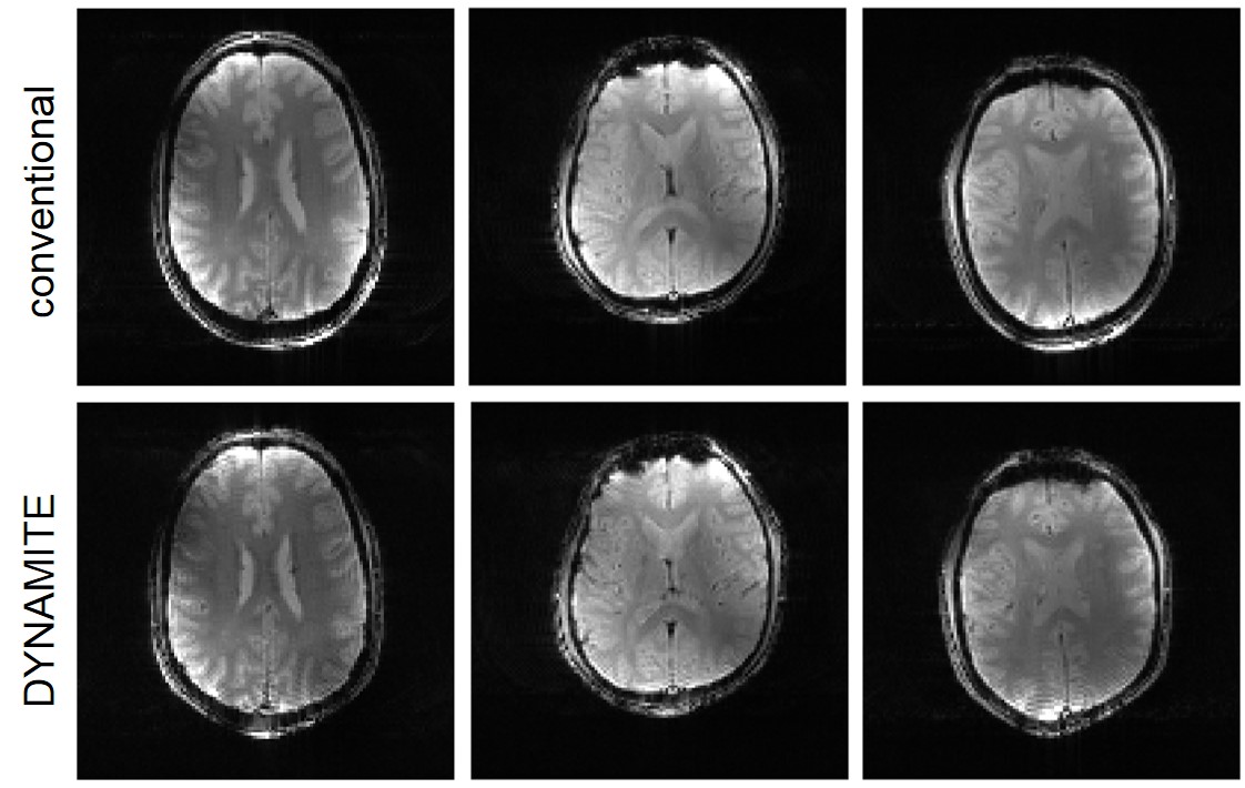
Quality assurance checks of the established multi-coil hardware (figure 1A) included the synthesis of well-defined, dynamically cycled field patterns such as a 100 Hz/cm linear field gradient that was rotated by 45-degree increments between slices (figure 1B). High accuracy of both shape and amplitude of the slice-specific gradient fields confirmed the general design and functionality of the derived multi-coil hardware and computational infrastructure (figure 1C). Our goal in this proof-of-principle research was to provide a physical replica of standard MRI sequences with DYNAMITE and to demonstrate full MRI capability in the human head at image quality comparable to conventional MR systems. To this end, all gradient patterns for slice-selection and in-plane phase and frequency encoding of conventional gradient-echo (figure 2A) and a spin-echo (not shown) sequences were replaced piece-by-piece with DYNAMITE-generated fields. The DYNAMITE gradient shapes were then played out by a series of well-timed TTL trigger signals to resemble the full MRI sequence (figure 2B). The resultant multi-slice spin-echo DYNAMITE images acquired from the human brain resembled those acquired with conventional gradient hardware throughout the FOV (figure 3). Similarly, high-level DYNAMITE MRI was achieved with both spin-echo (figure 4) and gradient-echo (figure 5) encoding from multiple subjects. Minor differences between conventional and DYNAMITE MRI can be explained by imperfectly calibrated MC magnetic field maps and will be addressed in the future by performing a higher-precision calibration over an extended phantom. Notably, the established sequence implementation allowed the replacement of individual sequence components (such as slice selection or in-plane encoding) and equivalent MRI results were obtained for all these scenarios (data not shown). The correspondence of conventional and DYNAMITE-encoded MRI strongly implies that the hundreds of gradient fields dynamically generated by DYNAMITE indeed closely resembled those provided by conventional gradient coils.CONCLUSIONS
The first successful realization of DYNAMITE MRI of the in vivo human brain at image fidelity comparable to MRI using conventional gradient coils is expected to pave the way for full-fledged human DYNAMITE MRI systems.Acknowledgements
This research was supported by NIH grants R24-MH105998, R01-EB014861, T32-EB008389 and U01-EB025153.References
1. Juchem, C., T.W. Nixon, S. McIntyre, D.L. Rothman, and R.A. de Graaf, Magnetic field modeling with a set of individual localized coils. J Magn Reson 204:281-9 (2010).
2. Juchem, C., D. Green, and R.A. de Graaf, Multi-coil magnetic field modeling. J Magn Reson 236:95-104 (2013).
3. Juchem, C., O.M. Nahhass, T.W. Nixon, and R.A. de Graaf, Multi-slice MRI with the dynamic multi-coil technique. NMR Biomed 28:1526-34 (2015).
4. Rudrapatna, S.U., F. Fluerenbrock, W.T. Nixon, R.A. de Graaf, and C. Juchem, Combined Imaging and Shimming with the Dynamic Multi-Coil Technique. Magn Reson Med (2018), epub ahead of print.
5. Juchem, C., T.W. Nixon, and R.A. de Graaf. Multi-Coil Imaging with Algebraic Reconstruction. Proc. ISMRM 2012, p.2545.
6. Juchem, C. B0DETOX - B0 Detoxification Software for Magnetic Field Shimming. Columbia TechVenture (CTV), License CU17326 (2017). Available from: innovation.columbia.edu/technologies/cu17326_b0detox.
7. Nixon, T.W., S. McIntyre, and R.A. de Graaf. The Design and Implementation of a 64 Channel Arbitrary Gradient Waveform Controller. Proc. ISMRM 2017, p.0969.
8. Adriany, G., P.F. Van de Moortele, F. Wiesinger, S. Moeller, J.P. Strupp, P. Andersen, C. Snyder, X. Zhang, W. Chen, K.P. Pruessmann, P. Boesiger, T. Vaughan, and K. Ugurbil, Transmit and receive transmission line arrays for 7 Tesla parallel imaging. Magn Reson Med 53:434-45 (2005).
Figures