0210
Simultaneous Measurement of functional MRI and MRS by Fast Non-water Suppressed Keyhole MR Spectroscopy Imaging1Weldon School of Biomedical Engineering, Purdue University, West Lafayette, IN, United States, 2School of Health Sciences, Purdue University, West Lafayette, IN, United States, 3Department of Psychiatry, McGill University, Montreal, QC, Canada, 4Centre d'Imagerie Cérébrale, Douglas Mental Health University Institute, Montreal, QC, Canada, 5Wellcome Centre for Integrative Neuroimaging, University of Oxford, Oxford, United Kingdom, 6Department of Radiology and Imaging Sciences, Indiana University School of Medicine, Indianapolis, IN, United States
Synopsis
The non-water suppressed magnetic resonance spectroscopic imaging (MRSI) sequence with concentric k-space trajectory was proposed to measure functional MRI and MRSI signals simultaneously. A right-hand finger-tapping task was performed at 3T MRI scanner to test the simultaneous hemodynamic and neurochemical measurements at human primary motor cortex. The results showed a significant overlap between T2* and metabolite (glutamate) changes.
Introduction
Combined fMRI-MRS is a novel method to non-invasively investigate functional activation in the human brain using simultaneous acquisition of hemodynamic and neurochemical measures (1). However, current methods typically consist of single voxel MRS with dimensions ranging from 8 mm3 to 27 mm3, and thus lack anatomical specificity. Magnetic resonance spectroscopic imaging (MRSI) allows neurochemical profiles to be acquired from multiple voxels simultaneously over large regions of the brain. Simultaneous acquisition of water and metabolite images utilizing metabolite-cycling MRSI provides the potential to collect functional MRI (fMRI) and MRSI (fMRSI) signals simultaneously (2). The aim of this project was to investigate the feasibility of utilizing a non-water suppressed MRSI sequence with concentric k-space trajectory (CRT) to perform fMRI and fMRSI simultaneously. To achieve this, we propose the use of keyhole imaging (3) (4), which has been used in dynamic imaging like functional MRI or cardiac functional examinations to reduce the acquisition time.Methods
The measurement was performed on a whole-body 3T MRI scanner (Siemens Healthineers, Erlangen, Germany). For the acquisition of a structural image a high-resolution T1-weighted MPRAGE sequence was used. A reference k-space for the keyhole scheme was acquired using metabolite-cycled DW-CRT semi-LASER with the following parameters: points-per-ring=64, temporal samples=512, resolution=5x5x10 mm3, Rings=24, FOV=240x240x10 mm3, TR=1350 ms, TE=32 ms, interleaves=4 (5). The simultaneous fMRI-fMRSI data was acquired using the keyhole method with 4 concentric rings at the central part of the k-space (Figure 1). During the fMRI-fMRS measurement, the subject performed repetitive tapping of the right-hand index finger with the following timing: four alternate periods of 1.8 min of motor activation and rest (ON–OFF–ON–OFF), preceded by a 1.8 min rest period (9 min total duration).
To obtain the fMRI-fMRSI images, a central part of the reference k-space was replaced by each set of dynamic keyhole data to yield and approximation of a complete k-space acquisition. NUFFT gridding was performed without using any post-hoc density compensation, since this is not required for DW-CRT data.
The T2* changes (BOLD response) during the activation were calculated using the unsuppressed water signal. Then, BOLD-response was analyzed using AFNI software. Metabolite spectra were subsequently calculated by subtracting the alternating FIDs. The LCModel was used to quantify the metabolite spectrum for each MRSI voxel (6).
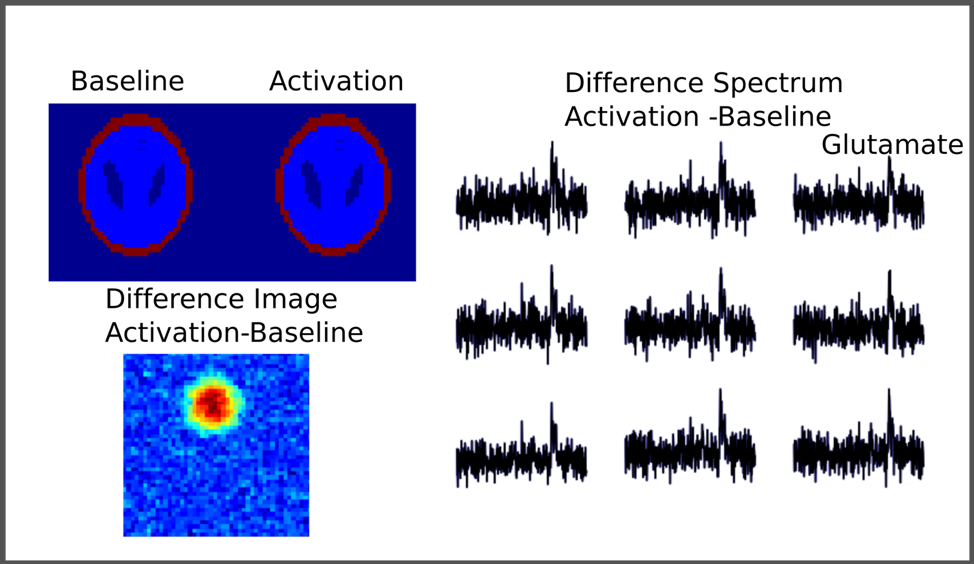
In addition to the in vivo experiment, a two-dimensional Shepp-Logan brain phantom was simulated to replicate the in vivo keyhole fMRI-fMRSI experiment. The spectra were simulated using GAMMA simulation with concentration values similar to previously reported in vivo values. To simulate functional activation, the Glutamate signal intensity of the spectra was increased by 4%, based on previously reported fMRS findings.
Results
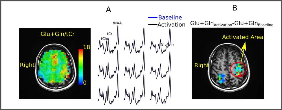
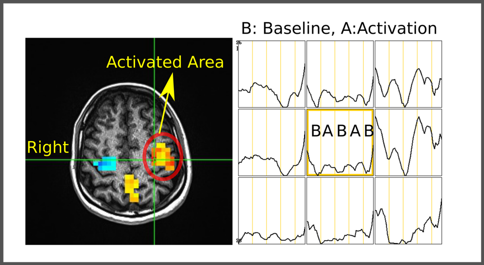
The simulation of keyhole MRSI acquisition results at two different glutamate levels (baseline and activation) demonstrate that the proposed method can detect 4% increase in Glutamate level by dynamically re-acquiring only the 4 concentric rings at the central part of the k-space (Figure 2). Figure 3a illustrates the reference Glu+Gln/tCr distribution of the first keyhole MRSI acquisition. In line with previous reports, Glu+Gln showed higher concentrations in the region of grey matter (GM) compared with white matter (WM). Results of LCModel analysis performed on metabolite spectra collected during baseline or stimulation periods were illustrated in Figure 3b. For each voxel, baseline concentrations were subtracted from activation concentrations. Figure 4 shows the statistical parametric map together with time series of T2* changes during the finger tapping. Preliminary analysis indicated a significant overlap in activation maps of T2* and metabolite changes.Conclusion
We provide the first spatial map of spatiotemporal interplay between dynamic glutamate changes and BOLD response in the human primary motor cortex during functional activation in response to finger tapping. These results represent a first step towards disambiguating contributions of cortical excitation and inhibition to stimulus evoked hemodynamic response.Acknowledgements
Supported by the Indiana CTSI, funded in part by grant #UL1TR001108 from the NIH, NCATS, CTS AwardReferences
1. Ip IB, Berrington A, Hess AT, Parker AJ, Emir UE, Bridge H. Combined fMRI-MRS acquires simultaneous glutamate and BOLD-fMRI signals in the human brain. Neuroimage 2017;155:113-119.
2. Emir UE, Burns B, Chiew M, Jezzard P, Thomas MA. Non-water-suppressed short-echo-time magnetic resonance spectroscopic imaging using a concentric ring k-space trajectory. NMR Biomed 2017;30(7).
3. van Vaals JJ, Brummer ME, Dixon WT, et al. "Keyhole" method for accelerating imaging of contrast agent uptake. J Magn Reson Imaging 1993;3(4):671-675.
4. Gao JH, Xiong J, Lai S, et al. Improving the temporal resolution of functional MR imaging using keyhole techniques. Magn Reson Med 1996;35(6):854-860.
5. Steel A, Chiew M, Jezzard P, et al. Metabolite-cycled density-weighted concentric rings k-space trajectory (DW-CRT) enables high-resolution 1 H magnetic resonance spectroscopic imaging at 3-Tesla. Sci Rep 2018;8(1):7792.
6. Provencher SW. Automatic quantitation of localized in vivo 1H spectra with LCModel. NMR Biomed 2001;14(4):260-264.
Figures