0197
Cystathionine as a marker for 1p/19q codeleted gliomas by in vivo magnetic resonance spectroscopy1Centre de NeuroImagerie de Recherche (CENIR), Institut du Cerveau et de la Moelle épinère (ICM), Paris, France, 2Sorbonne Université, UMR S 1127, Inserm U 1127, CNRS UMR 7225, Paris, France, 3Centre de Référence des Maladies Métaboliques, Service de Biochimie Métabolique, Hôpital Necker and Université Paris Descartes, Paris, France, 4AP-HP, Hôpital de la Pitié-Salpêtrière, Service de Neurologie 2, Paris, France, 5Department of Neurology, Foch Hospital, Suresnes, Paris, France, 6Center for Magnetic Resonance Research and Department of Radiology, University of Minnesota, Minneapolis, MN, United States, 7Onconeurotek tumor bank, Institut du Cerveau et de la Moelle épinère (ICM), Paris, France
Synopsis
Molecular markers such as mutation in isocitrate dehydrogenase (IDH) and codeletion of chromosome arms 1p and 19q (1p/19q codeletion) have highly benefited diagnosis and prognosis in brain gliomas. However, the biological effects of 1p/19q codeletion are still not clear. We report selective accumulation of cystathionine in IDH-mutated, 1p/19q codeleted gliomas observed by edited 1H magnetic resonance spectroscopy. Noninvasive detection of cystathione enables identification of glioma subtypes in vivo and opens up the possibility of investigating nonivasively cancer-specific metabolic pathways.
Introduction
Gliomas form a very heterogeneous group of malignant brain tumors1. The choice of appropriate tailored therapies is crucial for patients’ outcome, but requires precise identification of glioma subtype, which cannot be achieved by histology alone. The diagnostic and prognostic stratification of lower-grade brain gliomas highly benefits from the identification of molecular markers such as mutations in isocitrate dehydrogenase (IDH)2,3 and codeletion of chromosome arms 1p and 19q (1p/19q codeletion)4,5, both recognized as favorable predictive factors. Despite its high clinical relevance, the biological effects of 1p/19q codeletion are still not clear. We investigated the effect of 1p/19q codeletion on cancer-cells metabolism by combining in vivo 1H magnetic resonance spectroscopy (MRS) measurements in human brain gliomas with the analysis of a series of standard amino acids by liquid chromatography-mass spectroscopy (LC-MS) in human glioma biopsies.Methods
Sixty-five subjects with low-grade glioma were included in the study: 31 subjects (17 males; median age: 44 years) underwent the MRI/MRS examination and 47 brain tumor tissue samples (26 males; median age: 45 years) were analyzed with LC-MS. Acquisitions were performed using a 3 T whole-body Siemens system equipped with a 32-channel receive-only head coil. 3D FLAIR images were acquired to position the spectroscopic volume of interest (VOI) in the glioma (Figure 1). MR spectra were acquired with a single-voxel MEGA-PRESS6 sequence (TR = 2 s, TE = 68 ms) using previously described procedures and parameters7. Unsuppressed water scans were acquired from the same VOI for metabolite quantification and eddy current corrections using the same parameters as water suppressed spectra. For 8 subjects, the MEGA-PRESS acquisitions were performed also in the contralateral region outside the visible lesions. Single-shot spectra were frequency and phase aligned using the total choline signal at 3.22 ppm. All spectra were analyzed using LCModel8 with a simulated basis set including 2-hydroxyglutarate, cystathionine, γ-aminobutyric acid, glutamate, glutamine, glutathione, N-acetylaspartate, N-acetylaspartylglutamate, and the experimentally measured macromolecular spectrum. Automated immunohistochemical analysis (IHC) of IDH1 R132H and mutational status of IDH1 and IDH2 were determined as previously described9. The presence of 1p/19q codeletion was assessed by copy number variation analysis from next-generation sequencing targeted gene capture. Amino acid concentrations were measured with liquid chromatography coupled to tandem mass spectrometry (UPLC-MS/MS). For each tissue sample, amino acid concentrations were normalized to the median of amino acid concentrations, after exclusion of highly variable metabolites and those metabolites sometimes not reported in literature10.Results
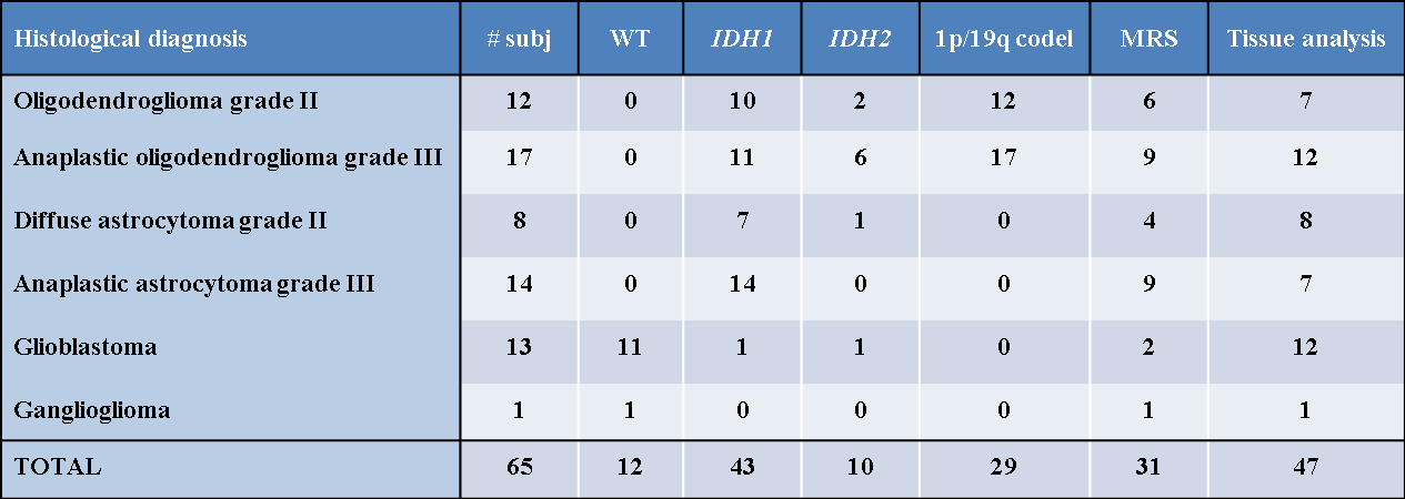
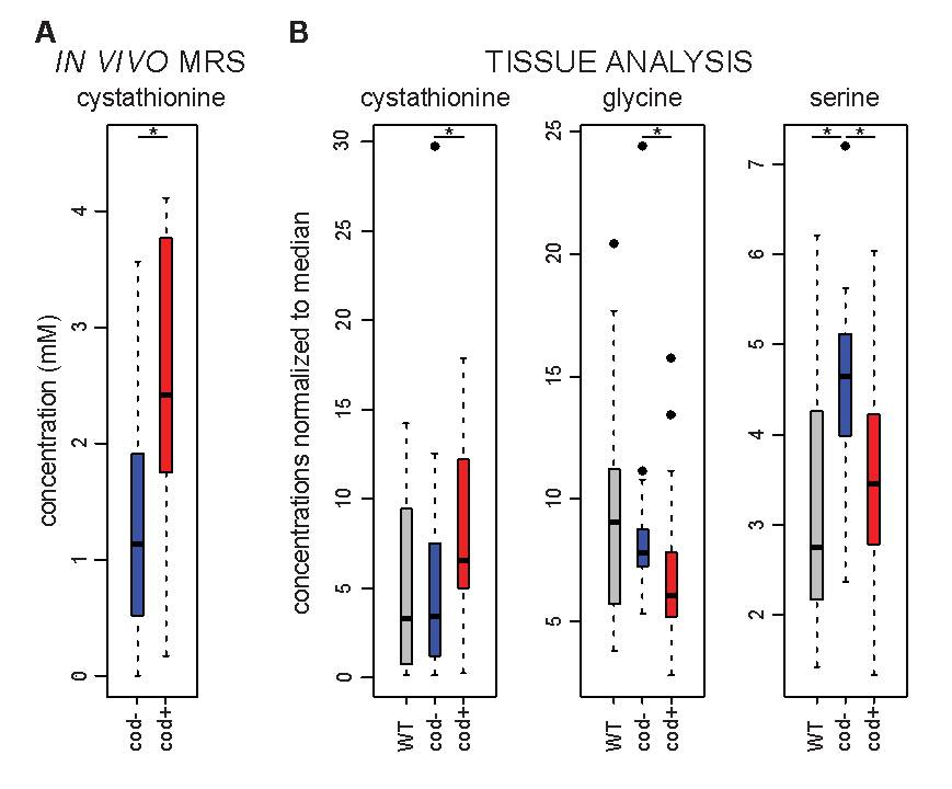
Integrated diagnosis according to the World Health Organization (WHO) 201611 are reported for the entire cohort in Table 1. In vivo spectra acquired in IDH1-mutated, 1p/19q codeleted gliomas are shown in Figure 1. Statistically significant higher cystathionine levels (two-tailed Student’s t-test: p = 0.014, Figure 2A) were detected in 1p/19q codeleted gliomas (average [cystathionine] = 2.6 mM; range: 0.2 - 4.1 mM; average CRLB = 36%), compared with IDH-mutated non-codeleted gliomas (average [cystathionine] = 1.25 mM; range: 0 - 3.6 mM; average CRLB = 235%). Cystathionine was visible in the MR spectra and measured reliably for [cystathionine] > 1.5 mM and CRLB < 30% in 12 of 15 subjects (80%) harboring the 1p/19q codeletion (Figure 1). Conversely, cystathionine was detectable in only 6 of the 14 subjects (43%) with IDH-mutated non-codeleted gliomas. Cystathionine was not detected in the healthy tissue. No significant differences in the other metabolites quantified by MRS were observed between codeleted and non-codeleted gliomas. Tissue analysis by LC-MS confirmed that cystathionine was significantly higher in IDH-mutated codeleted samples compared to their non-codeleted counterparts (Wilcoxon’s test: p = 0.048) (Figure 2B). In contrast, significantly lower levels of glycine and serine, both involved, as cystathionine, in glutathione biosynthesis pathway, were observed by LC-MS in codeleted versus non-codeleted gliomas (p = 0.023 and p = 0.005, respectively) (Figure 2B).Discussion and conclusion
Cystathionine is an immediate precursor of cysteine and glutathione in the transsulfuration pathway12. Our observation of higher, detectable cystathionine levels in IDH-mutated, 1p/19q-codeleted subjects in vivo agreed with tissue analysis, which additionally suggested a mechanism of pathology with possible therapeutic implications. In a few subjects, high cystathionine levels were observed without codeletion, yet they may have had undetected intragenic mutations (or epigenetic modifications). Cystathionine accumulation and other abnormal levels of amino acids in 1p/19q-codeleted gliomas (Figure 2) may result from partial genetic loss of two genes located on chromosome 1p: phosphoglycerate dehydrogenase (PHGDH) and cystathionine gamma-lyase (CTH), both involved in serine- and glutathione-pathways. Our results point to a possible selective vulnerability to serine and glutathione depletion in 1p/19q codeleted gliomas and suggest in vivo cystathionine as a candidate noninvasive marker to monitor cancer treatments in patients harboring these tumors.Acknowledgements
FB and SL acknowledge support from Investissements d’avenir [grant number ANR-10-IAIHU-06 and ANR-11-INBS-0006]. DD and MM acknowledge support from following National Institutes of Health grants: BTRC P41 EB015894 and P30 NS076408. The authors would like to thank Edward J. Auerbach, Ph.D., for implementing MRS sequences on the Siemens platform.References
1. Picca A, Berzero G, Sanson M. Current therapeutic approaches to diffuse grade II and III gliomas. Ther Adv Neurol Disord. 2018; 11:1-13.
2. Parsons DW, Jones S, Zhang X, et al. An integrated genomic analysis of human glioblastoma multiforme. Science. 2008; 321(5897):1807-1812.
3. Dang L, White DW, Gross S, et al. Cancer-associated IDH1 mutations produce 2-hydroxyglutarate. Nature. 2009; 462(7274):739-744.
4. Reifenberger J, Liu L, James CD, Wechsler W. Molecular Genetic Analysis of Oligodendroglial Tumors Shows Preferential Allelic Deletions on 19q and 1p. Am J Pathol. 1994; 145(5):16.
5. Idbaih A, Marie Y, Pierron G, et al. Two types of chromosome 1p losses with opposite significance in gliomas. Ann Neurol. 2005; 58(3):483-487.
6. Mescher M, Merkle H, Kirsch J, Garwood M, Gruetter R. Simultaneous in vivo spectral editing and water suppression. NMR Biomed. 1998; 11:266-272.
7. Branzoli F, Di Stefano AL, Capelle L, et al. Highly specific determination of IDH status using edited in vivo magnetic resonance spectroscopy. Neuro-Oncol. 2018; 20(7):907-916.
8. Provencher SW. Estimation of metabolite concentrations from localized in vivo proton NMR spectra. Magn Reson Med. 1993; 30(6):672-679.
9. Sanson M, Marie Y, Paris S, et al. Isocitrate dehydrogenase 1 codon 132 mutation is an important prognostic biomarker in gliomas. J Clin Oncol. 2009; 27(25):4150-4154.
10. Lefauconnier J-M, Portemer C, Chatagner F. Free amino acids and related substances in human glial tumours and in fetal brain: comparison with normal adult brain. Brain Res 1976; 117(1):105-113.
11. Louis DN, Perry A, Reifenberger G, et al. The 2016 World Health Organization classification of tumors of the central nervous system: a summary. Acta Neuropathol. 2016; 131(6):803-820.
12. Vitvitsky V, Thomas M, Ghorpade A et al. A Functional Transsulfuration Pathway in the Brain Links to Glutathione Homeostasis. J Biol Chem. 2006;281(47):35785–35793.
Figures
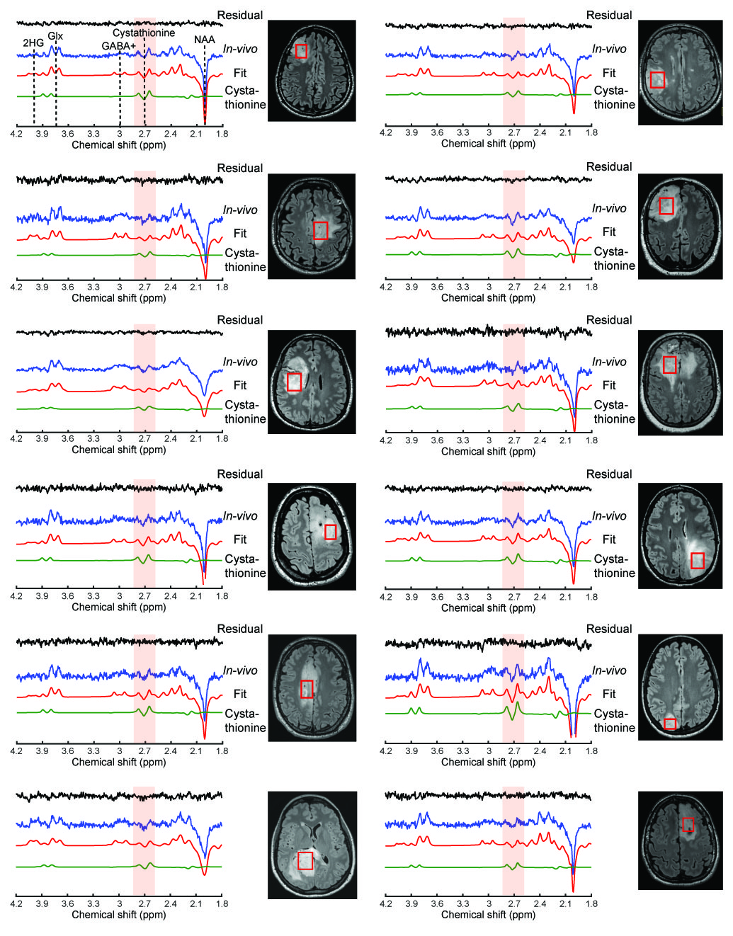
Figure 1. In vivo spectra acquired with edited spectroscopy at 3 T (blue lines) in the IDH1-mutated gliomas with 1p/19q codeletion are shown together with LCModel fits (red lines), the cystathionine contribution (green lines), and residuals (black lines). The location and size of the VOIs are shown on FLAIR images. The cystathionine pattern is visible at 2.7 ppm. Spectra are scaled with respect to the water signal. No line broadening was applied. NAA: N-acetylaspartate; GABA+: GABA + macromolecules; Glx: glutamate + glutamine; 2HG: 2-hydroxy glutarate.