0188
PET derived Raphe Nuclei [11C]-CUMI binding potential is associated with MR derived diffusion entropy in the frontal and temporal lobes1Biomedical Engineering, Stony Brook University, Stony Brook, NY, United States, 2Psychiatry, Stony Brook Medicine, Stony Brook, NY, United States, 3Radiology, Stony Brook Radiology, Stony Brook, NY, United States
Synopsis
Using a new DTI processing technique, named diffusion entropy, and dynamic PET data we observed a statistically significant inverse correlation between diffusion entropy and [11C]-CUMI binding potential (BPF) in a dataset with bipolar, major depressive disorder patients and controls. This newfound analysis can give rise to further understanding the pathophysiology that undermines psychiatric diseases and, also, a non-invasive, non-ionizing radiation procedure to estimate [11C]-CUMI BPF.
Introduction
Serotonergic dysfunction is implicated in the current understanding of virtually all mood disorders1. The raphe nuclei (RN) are the preeminent source of serotonin for higher brain regions2. A rich system of projections originating from the RN supplies ascending serotonergic innervation, most interestingly to the frontal and temporal lobe areas widely implicated in mood disorders3. 5-HT1A is among the most expressed serotonin receptors in the brain, with pronounced density in the cortical matter and RN4; wherein RN 5-HT1A binding has served as a moderating factor in treatment response5-6.
Recent work suggests that serotonergic transmission might mediate abnormal gray matter morphology observed in several disorders. Most recently, Pillai et al. demonstrated a correlation between cortical thickness throughout the brain and PET-derived 5-HT1A density in the RN7. We have recently observed a novel biomarker in individuals with bipolar depression using a new DTI processing technique named diffusion entropy, in which we not only observed to significantly differ between patients with bipolar depression and matched controls, but also capable of predicting diagnostic status. Therefore, we hypothesize that diffusion entropy is related to RN 5-HT1A binding, which might allow us to have an MR derived biomarker for PET parameters.
Methods
Our dataset consisted of 59 pairs of PET and processed diffusion tensor imaging (DTI) MRI data. Specifically, this population comprised 28 healthy controls, 19 subjects BD with and 12 subjects with MDD. Dynamic PET data were collected following the injection of a single bolus of up to 5mCi of [11C]-CUMI. MD entropy was calculated for the frontal and temporal lobe using our previously published technique8. Summarily, for each subject, voxel-wise MD values in the lobes were grouped into 5 histogram bins, from which MD entropy was calculated for the entire ROI by the following formula.
$$H=∑P(x_i)log(P(x_i))$$
Where P(xi) denotes the height of the histogram bin centered at xi. All statistical analysis was performed using SPSS. Spearman partial correlation between [11C]-CUMI BPF in the raphe nuclei and MD entropy in the frontal lobe, controlling for age, sex and scan site were conducted.
Results
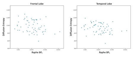
Scatter plots of frontal and temporal lobe diffusion entropy as a function of RN [11C]-CUMI BPF, without any demographic corrections, are shown in Figure 1. In both instances, we observe a statistically significant inverse correlation (pFrontal=0.045; pTemporal=0.026), although these measurements do not maintain statistical significance when adjusting for the two comparisons made using Bonferroni correction.
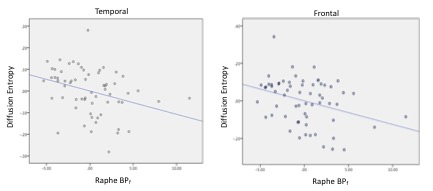
Following this assessment of uncorrected values, we investigated the confounding effects of age, sex and MRI and PET scan site on the data. Partial correlation with these effects corrected strengthened both the magnitude and significance of the correlation in both instances (pFrontal=0.007; pTemporal = 0.015). Figure 2 provides the resultant scatter plots of this analysis. Finally, we controlled for the effects of age, sex, scan site and subject diagnostic status. This correction led to largely similar observations as the previous correction (rFrontal=-.352 pFrontal=0.002; rTemporal=-0.388 pTemporal=0.003).
We sought to investigate whether the observed correlations between [11C]-CUMI BPF of the midbrain and frontotemporal diffusion entropy were unique to the raphe nuclei. Excluding the midbrain, regression analysis with all previously applied corrections against 50 additional PET regions of interest resulted in no observations with p<0.05.
Discussion
In the present hypothesis-driven study, we used PET and MRI and observed a significant inverse correlation between RN 5-HT1A binding and diffusion entropy metric. Diffusion entropy is a measure of the distribution of mean diffusivity values in a region, with low diffusion entropy indicating relative homogenous MD values and high diffusion entropy indicating heterogeneously distributed values. 5-HT1A is among the most expressed serotonin receptors throughout the brain, with pronounced density in the cortical matter and RN. Therefore, the effect that we may have observed may initially be viewed as non-specific to the RN. Yet, we prove that this effect is specific to the raphe since our regression analysis resulted in only one region with significant finding.Conclusion
This analysis observed that frontotemporal diffusion entropy to be significantly inversely correlated with raphe [11C]-CUMI BPF. We hope to use this MR derived metric to further understand the pathophysiology that undermines complex diseases such as depression. Diffusion entropy may be an indicator for astrocyte microstructural organization since a large amount of evidence labels astrocytic dysfunction as a possible cause for depression9-11, yet further research is needed. Since the diffusion entropy metric is found solely through MRI techniques, this may allow estimating 5HT1A BPF a non-invasive, non-ionizing radiation procedure.Acknowledgements
No acknowledgement found.References
1. K. A. Michelsen, J. Prickaerts and H. W. Steinbusch, Progress in brain research 172, 233-264 (2008).
2. B. L. Jacobs and E. C. Azmitia, Physiological reviews 72 (1), 165-229 (1992).
3. J. Kaufman, C. DeLorenzo, S. Choudhury and R. V. Parsey, European Neuropsychopharmacology 26 (3), 397-410 (2016).
4. H. Hall, C. Lundkvist, C. Halldin, L. Farde, V. Pike, J. McCarron, A. Fletcher, I. Cliffe, T. Barf and H. Wikström, Brain research 745 (1-2), 96-108 (1997).
5. R. V. Parsey, D. M. Olvet, M. A. Oquendo, Y.-y. Huang, R. T. Ogden and J. J. Mann, Neuropsychopharmacology 31 (8), 1745-1749 (2006).
6. M. J. Lan, N. Hesselgrave, A. Ciarleglio, R. T. Ogden, G. M. Sullivan, J. J. Mann and R. V. Parsey, Synapse 67 (11), 773-778 (2013).
7. R. L. Pillai, A. Malhotra, D. D. Rupert, B. Weschler, J. C. Williams, M. Zhang, J. Yang, J. J. Mann, M. A. Oquendo and R. V. Parsey, Human brain mapping (2018).
8. K. Spuhler, E. Bartlett, J. Ding, C. DeLorenzo, R. Parsley and C. Huang, (Synapse, 2017).
9. G. Rajkowska, J. J. Miguel-Hidalgo, J. Wei, G. Dilley, S. D. Pittman, H. Y. Meltzer, J. C. Overholser, B. L. Roth and C. A. Stockmeier, Biological psychiatry 45 (9), 1085-1098 (1999).
10. R. A. Gittins and P. J. Harrison, Journal of affective disorders 133 (1), 328-332 (2011).
11. M. P. Bowley, W. C. Drevets, D. Öngür and J. L. Price, Biological psychiatry 52 (5), 404-412 (2002).
Figures