0174
An MRS investigation of superior visual search abilities in children with autism spectrum disorder: Evidence for enhanced top-down attentional filteringDavid A Edmondson1,2, Pingyu Xia1, Brandon Keehn3, and Ulrike Dydak1,2
1School of Health Sciences, Purdue University, West Lafayette, IN, United States, 2Department of Radiology and Imaging Sciences, Indiana University School of Medicine, Indianapolis, IN, United States, 3Department of Speech, Language, & Hearing Sciences, Purdue University, West Lafayette, IN, United States
Synopsis
Processing strengths or “islets of ability” associated with autism spectrum disorder (ASD), especially in visual search, have been of continued interest as they provide insight into how those with ASD perceive the world around them. In typically-developing (TD) individuals, region-specific concentrations of GABA are associated with differences in attention and perception. ASD may be associated with an excitatory-inhibitory imbalance, however, it remains unclear how this may contribute to superior search abilities. To test this, 37 ASD and TD children participated in a magnetic resonance spectroscopy study using MEGA-semi-LASER to detect GABA concentrations in cortical regions associated with attention and perception.
Introduction
Individuals with autism spectrum disorder (ASD) excel at visual search, performing better than their typically-developing (TD) peers across the lifespan1. Evidence of augmented ASD search superiority with increasing target-distractor similarity has been used to support the hypothesis that enhanced perceptual functioning contributes to faster search. Others have suggested that faster search may be due to differences in the distribution of attention and attentional filtering rather than enhanced lower-level perceptual processing. Thus, the precise mechanism underlying superior search and the brain bases for this advantage remains unknown. One model has hypothesized that ASD is associated with an imbalance of glutamatergic and GABAergic signaling.2 In addition, inter-individual differences in attention and perception in TD individuals are associated with region-specific concentrations of GABA3,4 However, the contributions of atypical GABAergic function to enhanced visual search abilities in ASD has not been determined. To test this, we used magnetic resonance spectroscopy (MRS) to measure GABA using MEGA-semi-LASER in the bilateral visual cortex (VIS), right temporal parietal junction (TPJ), and right frontal eye fields (FEF).Methods
Twenty-three children with high-functioning ASD (19 males, 4 females, Average Age = 11.7 years, SD = 1.2) and 14 TD children (10 males, 4 females, Average Age = 11.7, SD = 1.5) participated. Participants completed multiple experimental paradigms prior to scanning including a visual search task (VST)5. Participants’ task was to indicate the presence or absence of a target (vertical line) embedded within arrays of distractors (tilted lines) that varied in set size (18, 24, 36 items) (Figure 1a,b). Search speed was measured as reaction time (RT) to determine presence or absence of the target item. The slopes (a measure of search efficiency, reflecting the RT cost of each additional distractor) and y-intercepts (associated with non-search, perceptual processes) of the RT x set size functions were calculated for target present and absent conditions (Figure 1c). All scans were obtained on a 3T Siemens Prisma MR Scanner at Purdue University. MEGA-semi-LASER6,7 (TE = 68ms, TR = 2000ms, Averages = 128) was used to measure GABA, Glx, NAA and tCr (Cr + PCr) in the VIS (30mm x 30mm x 20mm), TPJ (30mm x 30mm x 20mm), and FEF (30mm x 30mm x 20mm)3 (Figure 2). Difference and Off spectra were quantified using LCModel8. Repeated measures ANOVAs were used to assess differences between groups and regions of interest. Pearson correlation tests and were used to assess relationships between metabolites and VST parameters (RT(present & absent), y-intercept(present & absent), and slope(present & absent)).Results
No group differences were found between ASD and TD in any region of interest (ROI) for GABA, Glx, and tCr. However, there was a significant main effect of group for NAA, with ASD having lower NAA than TD (F(1,94) = 10.94, p = 0.001). While NAA was, on average, lower for ASD than TD in all three ROIs, post-hoc Tukey test suggests that NAA was significantly lower (p < 0.01) in the TPJ but not for the FEF and VIS (Figure 3). No differences in RT(present or absent) or other VST measures were noted. In ASD, we found a negative association between GABA+ and slope(absent) (r = -0.47, p = 0.047) in the FEF (Figure 4) while, in the VIS, NAA was positively correlated with slope(absent) (r = 0.52, p = 0.02). No significant relationships were found in TD children between metabolites or VST.Discussion
GABA concentrations in the three ROIs were found to be similar in ASD versus TD, which supports findings in other studies. Nonetheless, in the FEF, we found that as GABA increases, VST performance improves for ASD, but not in TD, suggestive of improved attentional filtering. We also found NAA, commonly believed to be a neuronal marker for function, to be lower in ASD than TD across all three regions, but only significantly different in the TPJ, as has also been found by others9–11. This suggests that NAA, acting as a marker of neuronal function, may be indicating a difference in GABAergic signaling efficiency in ASD due to neuronal dysfunction.Conclusion
Together, our findings are similar to prior reports, but suggest that autistic visual search superiority may be due to differences in GABA signaling in the FEF. These differences may be the underlying cause of improved visual search via improved attentional filtering. Decreased NAA in ASD compared to TD may be linked to dysfunctional GABA signaling, as has been found in animal models12. This may explain why average cortical GABA levels do not differ between ASD and TD, but VST performance improves in relation to increased GABA levels in ASD.Acknowledgements
This study was supported by a Purdue Neuroscience Pilot Grant and NIEHS F31 ES028081.References
- Kaldy Z, Giserman I, Carter AS, Blaser E. The Mechanisms Underlying the ASD Advantage in Visual Search. J Autism Dev Disord. 2016;46(5):1513-1527. doi:10.1007/s10803-013-1957-x.
- Hussman JP. Suppressed GABAergic Inhibition as a Common Factor in Suspected Etiologies of Autism. J Autism Dev Disord. 2001;31(2):247-248. doi:10.1016/0002-9610(92)90118-B.
- Sumner P, Edden RAE, Bompas A, Evans CJ, Singh KD. More GABA, less distraction: a neurochemical predictor of motor decision speed. Nat Neurosci. 2010;13(7):825-827. doi:10.1038/nn.2559.
- Edden RAE, Muthukumaraswamy SD, Freeman TCA, Singh KD. Orientation Discrimination Performance Is Predicted by GABA Concentration and Gamma Oscillation Frequency in Human Primary Visual Cortex. J Neurosci. 2009;29(50):15721-15726. doi:10.1523/JNEUROSCI.4426-09.2009.
- Kemner C, Van Ewijk L, Van Engeland H, Hooge I. Brief report: Eye movements during visual search tasks indicate enhanced stimulus discriminability in subjects with PDD. J Autism Dev Disord. 2008;38(3):553-558. doi:10.1007/s10803-007-0406-0.
- Andreychenko A, Boer VO, Arteaga De Castro CS, Luijten PR, Klomp DWJ. Efficient spectral editing at 7 T: GABA detection with MEGA-sLASER. Magn Reson Med. 2012;68(4):1018-1025. doi:10.1002/mrm.24131.
- Marjańska M, Auerbach EJ, Valabrègue R, Van de Moortele P-F, Adriany G, Garwood M. Localized 1H NMR spectroscopy in different regions of human brain in vivo at 7 T: T2 relaxation times and concentrations of cerebral metabolites. NMR Biomed. 2012;25(2):332-339. doi:10.1002/nbm.1754.
- Provencher SW. Estimation of metabolite concentrations from localized in vivo proton NMR spectra. Magn Reson Med. 1993;30(6):672-679. doi:10.1002/mrm.1910300604.
- Kleinhans NM, Schweinsburg BC, Cohen DN, Müller R-A, Courchesne E. N-acetyl aspartate in autism spectrum disorders: regional effects and relationship to fMRI activation. Brain Res. 2007;1162:85-97. doi:10.1016/j.brainres.2007.04.081.
- Hardan AY, Fung LK, Frazier T, et al. A proton spectroscopy study of white matter in children with autism. Prog Neuro-Psychopharmacology Biol Psychiatry. 2015;66:48-53. doi:10.1016/j.pnpbp.2015.11.005.
- Carvalho AP, Violante IR, Mouga S, Oliveira G, Castelo-Branco M. Medial Frontal Lobe Neurochemistry in Autism Spectrum Disorder is Marked by Reduced N-Acetylaspartate and Unchanged Gamma-Aminobutyric Acid and Glutamate + Glutamine Levels. J Autism Dev Disord. 2018;48(5):1467-1482. doi:10.1007/s10803-017-3406-8.
- Horder J, Andersson M, Mendez MA, et al. GABAA receptor availability is not altered in adults with autism spectrum disorder or in mouse models. Sci Transl Med. 2018;10(461):eaam8434. doi:10.1126/scitranslmed.aam8434.
Figures
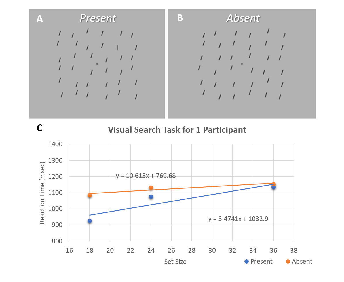
Figure 1: Visual Search Task. A
& B) Example of target present (A)
and target absent (B)
in a 36-item visual search array.
C) Reaction time
for each set is collected and averaged for set sizes of 18, 24, and 36. The
slope is calculated from a linear fit of RT to these three set sizes. The y-intercept is the baseline RT and is
from extrapolating the slope to a set size of 0.
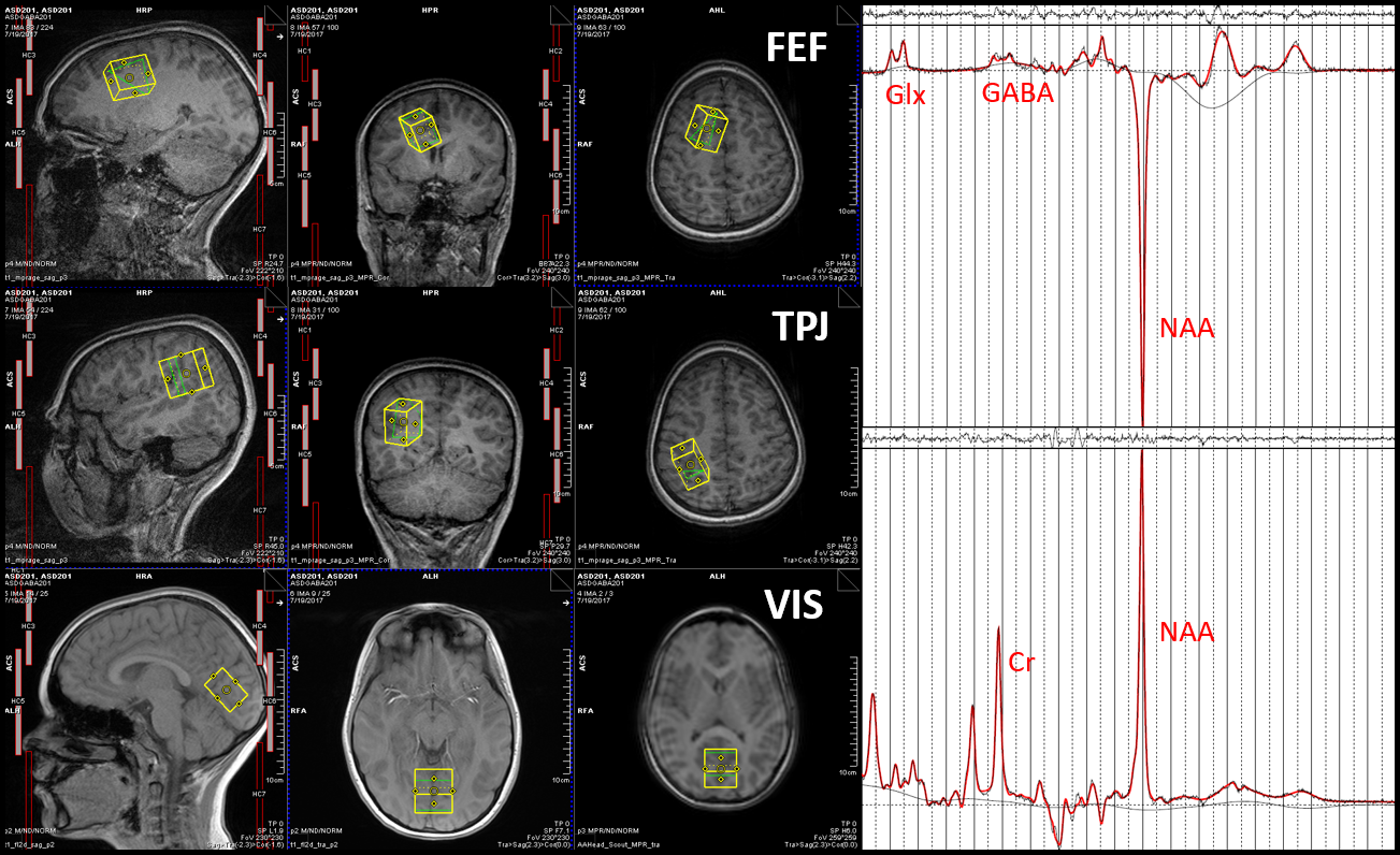
Figure 2: Voxel Placement and Example
MEGA-Semi-LASER spectra. A)
Voxel placement in the FEF, TPJ, and VIS in a participant. B) Difference
spectrum obtained using MEGA-Semi-LASER localization. C) Off Spectrum obtained
using MEGA-Semi-LASER localization
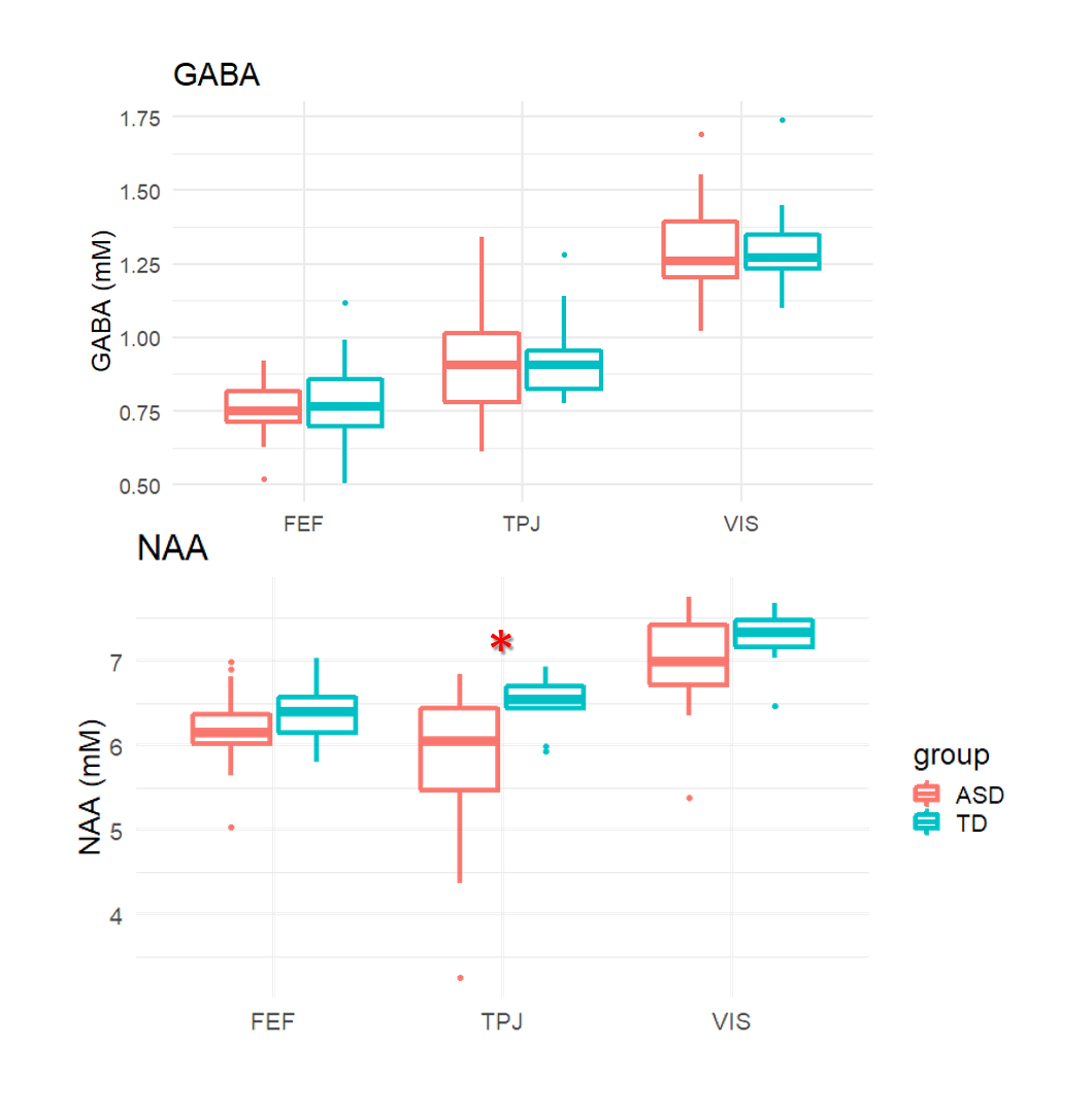
Figure 3: Group
differences in NAA and GABA between ASD and TD. There were no group differences in GABA, but
obvious regional differences between FEF, TPJ, and VIS. There was main effect
in NAA
with ASD having
lower NAA than
TD (F(1, 94) = 10.94, p = 0.001). Post hoc Tukey test found that there was
only a significant difference between TD and ASD in the TPJ (p = 0.01). There
were no group
differences in Glx or tCr (not
shown).
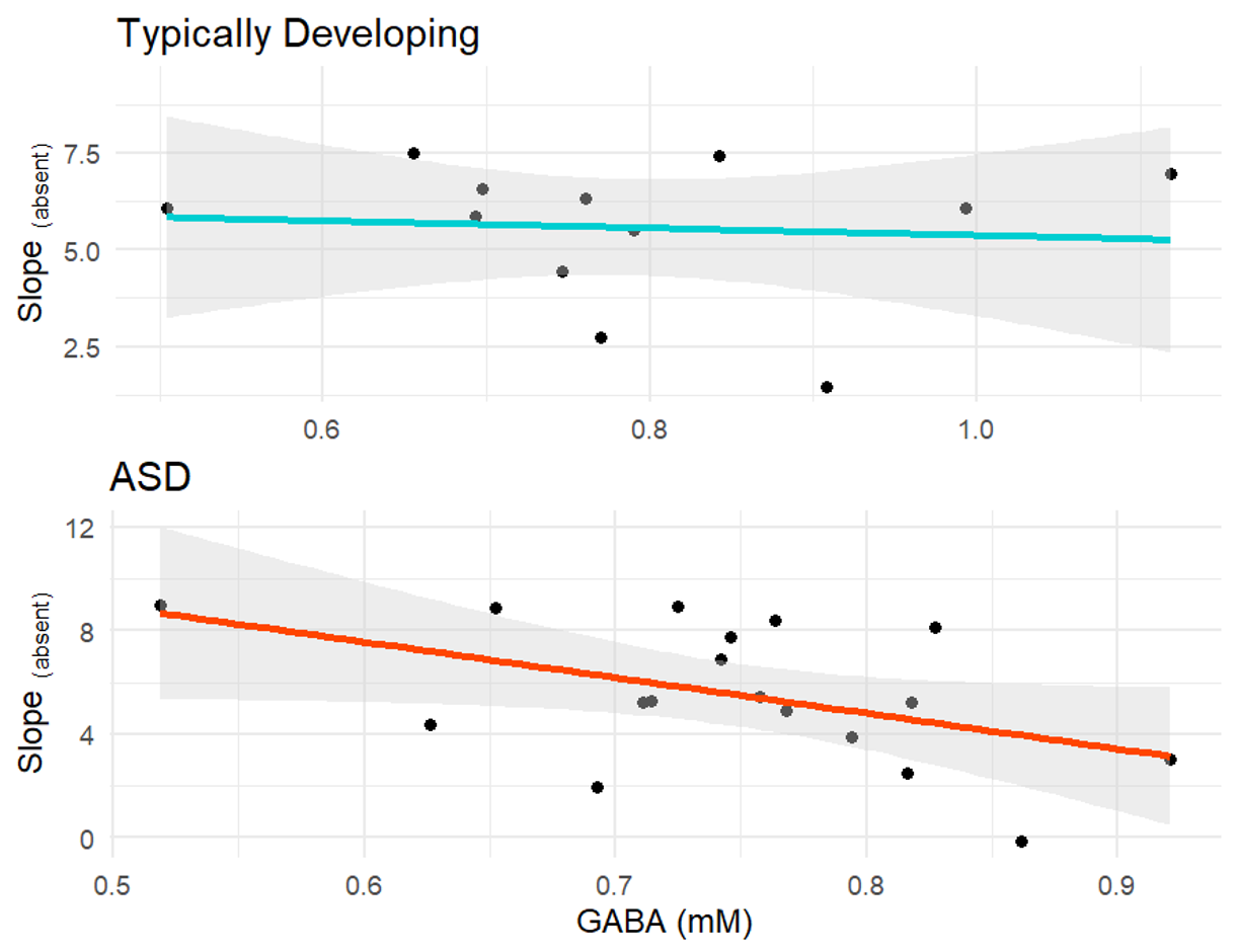
Figure 4: GABA versus Slope(absent) between ASD and TD. The linear
relationship between GABA and Slope(absent) is
different between ASD and TD. ASD has a significant correlation GABA+
& slope absent (r = -0.47 , p = 0.047 ) suggesting that increased GABA leads to
a decreased slope, thus better VST performance in the absent condition. This
relationship does not exist in TD.