0172
Antibiotic rifaximin for treatment of chronic liver disease-induced HE: a longitudinal in vivo 1H-MRS study of brain metabolism1LIFMET, Ecole Polytechnique Fédérale de Lausanne, Lausanne, Switzerland, 2CIBM, Ecole Polytechnique Fédérale de Lausanne, Lausanne, Switzerland, 3Service of Biomedecine, University Hospital of Lausanne, Lausanne, Switzerland, 4Swiss Center for Liver Disease in Children, Department of Pediatrics, University Hospitals Geneva, Geneva, Switzerland
Synopsis
Rifaximin
is a commonly-used antibiotic to treat hepatic encephalopathy(HE), a complex neuropsychiatric
syndrome caused by hepatic dysfunction.
Rifaximin reduces the production of gut ammonia, the main toxin in HE
pathogenesis. We hypothesized that the effect of rifaximin on
neurometabolic profile is dose-related. Therefore, in this study, the effects of rifaximin administered at 6x
human-dose were
assessed, in vivo and longitudinally
on brain metabolites in bile-duct ligated(BDL) rats using 1H-MRS at 9.4T,
biochemical and behavioral tests. They were compared with non-treated and
human-dose treated rats. We showed that higher-dose rifaximin treatment
was associated with positive effects on brain Gln,Glu and osmoregulation.
Introduction
Hepatic encephalopathy(HE) is a complex neuropsychiatric syndrome affecting the brain caused by hepatic dysfunction. Treatments for HE have focused on reducing plasma ammonia(NH4+) levels, considered to be the main toxin in HE pathogenesis. Rifaximin is a non–absorbable antibiotic which inhibits the division of colonic bacteria responsible for urea deamination, reducing the production of gut ammonia. In humans, rifaximin has been shown to reduce the frequency of HE episodes1, but the molecular mechanisms behind this effect are unknown.
In a previous study using bile duct ligated(BDL) rats, a model of chronic liver disease(CLD)2,3, we showed that rifaximin at human dose may help reduce brain Gln levels in early stages of HE4 (high Gln levels are the result of NH4+ detoxification in non-treated rats). These findings raised the question of the efficacy of the dose used, therefore we hypothesized that the effect of rifaximin on neurometabolic profile is dose-related.
To test this hypothesis, the effects of a higher dose of rifaximin (6x human-dose, resulting from the conversion from human dose to rat5) were assessed, in vivo and longitudinally on brain metabolites in BDL rats using 1H-MRS at 9.4T, biochemical and behavioral tests. The results were compared to BDL non-treated rats(n=17) and BDL rats treated with a human dose of rifaximin(n=12)4.
Methods
Adult Wistar rats(n=8) underwent bile duct ligation. Plasma measurements of NH4+, bilirubin and MRS-scans were performed before BDL(‘week0’) and at weeks 2,4,6,8 post-BDL. Rifaximin was administered orally twice daily (6x-human-dose=97.3mg/kg/day) starting 2 weeks after BDL-surgery(‘week2’).
In vivo 1H-MRS was performed on a 9.4T MRI system(Varian/Magnex Scientific) using a inhouse-built 14mm diameter quadrature 1H-coil as a transceiver. Metabolites evolution was studied in the hippocampus(2x2.8x2mm3), a region implicated in cognitive deficits in chronic HE, using SPECIAL6 sequence (TE=2.8ms, TR=4000ms, 160 averages). Field inhomogeneity was corrected using FASTESTMAP(linewidth=9-11Hz). Metabolite concentrations were estimated by LCModel using water as internal reference. Open field test was performed at week 4,6 and 8 to evaluate motor activity as a marker of HE severity7.
Results and Discussion
Plasma measurements of bilirubin confirmed the presence of CLD in both groups of rats. All rats displayed a similar ammonium increase regardless of the group(Fig.1).
The high quality of the spectra throughout the study allowed the quantification of 18 brain metabolites(Fig.2). Both groups exhibited a significant increase of brain Gln(p<0.001) due to ammonia detoxification, and a subsequent reduction of other brain osmolytes(Ins, Tau, tCho) as an osmolatory response(Fig.4).
Interestingly, 1H-MRS revealed some significant differences between the ‘high-dose rifaximin’ rats and non-treated rats: Gln increase was lower in the ‘high-dose rifaximin’ group at week6 and at week8, both in absolute value and relative to week2(+42% vs +118% at week8,Fig 3). Moreover, a decrease of Gln was observed between week4 and week6 in the ‘high-dose rifaximin’ group( ̴̴-10%) while it was continuously increasing in the non-treated group(Fig.2&Fig.3).
The neurotransmitter Glu showed no significant decrease in the treated group, whereas it reached -14% at week8 in the non-treated group(p<0.01,Fig.4). The diminution of the osmolyte Ins was also less marked in the ‘high-dose rifaximin’ group. Cr, a metabolite known for its role in energy metabolism and reported to participate in osmoregulation and neuroprotection8,9 showed a decrease in the non-treated group at week8( ̴̴-12%,p<0.01) and no decrease in the ’high-dose rifaximin’ group. The antioxidant Asc exhibited a small decrease in both groups at week8 and no significant variation in Lac were observed(Fig.4).
While rifaximin at human dose appeared to have an effect only at the early stages of the disease, a higher dose gave stronger positive effects on the neurometabolic profile(Fig.3), suggesting an effect of the treatment probably by decreasing the pool of deleterious bacteria in the gut. Importantly, no differences between the groups were observed in behavioural test performance and the ‘high-dose rifaximin’ rats had the tendency to move less. It is therefore possible that such a high-dose of antibiotics also leads to some noxious effects as antibiotics imbalance the intestinal microbiota, affect intestinal permeability10 and may worsen the general condition of the rats, e.g by promoting inflammatory response11.
Conclusion
In BDL rats, rifaximin treatment at higher dose was associated with positive effects on brain Gln,Glu and osmoregulation. The clinical implications of these findings are important given that rifaximin is a commonly used antibiotic in the treatment of HE. To our knowledge this is a first study comparing the effects of these two rifaximin doses on brain metabolites.
Moreover, in view of our results, we believe that high doses of antibiotics should be combined with probiotics to further improve the overall outcome (neurological/general condition), in line with our previous findings12,13.
Acknowledgements
Supported by CIBM of the UNIL, UNIGE, HUG, CHUV, EPFL, the Leenaards and Jeantet Foundations, and the SNSF project no 310030_173222/1.References
1Bajaj et al, Ap&T 2016 ; 2Biecker et al, J Pharmacol Exp Ther 2005; 3Butterworth et al, Liver Int 2009; 4Flatt et al, ISMRM 2017; 5Nair AB, Jacob S. J Basic Clin Pharma 2016; 6Mlynárik et al, Magn Reson Med 2006; 7 Leke et al, Plos one 2012; 8 Rae, NeurochemRes 2014; 9Braissant et al., MolGenetMetab 2010; 10Tulstrup et al, Plos one, 2015; 11Knoop KA et al, Gut. 2016; 12Rackayova et al, ISMRM 2016; 13Flatt et al, ISMRM 2018Figures
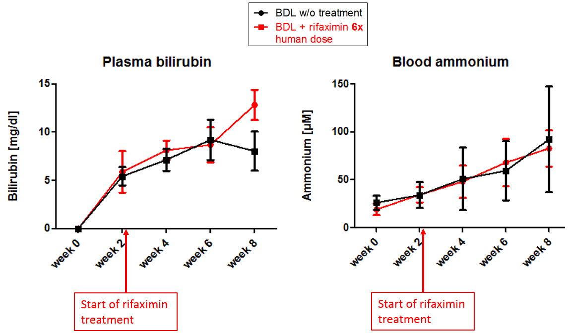
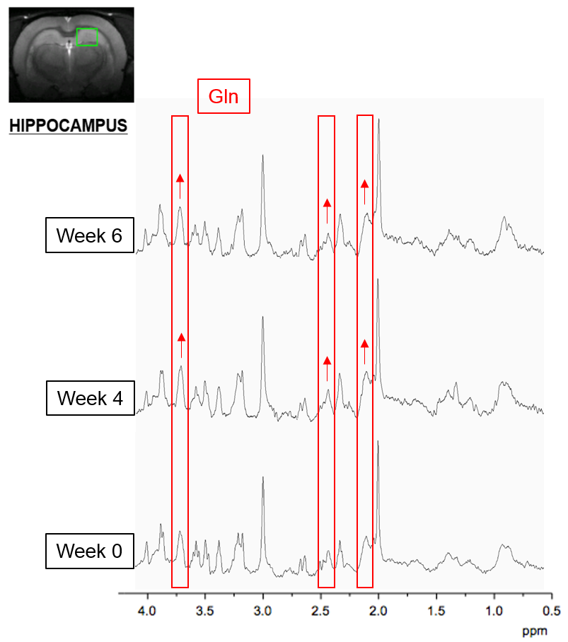
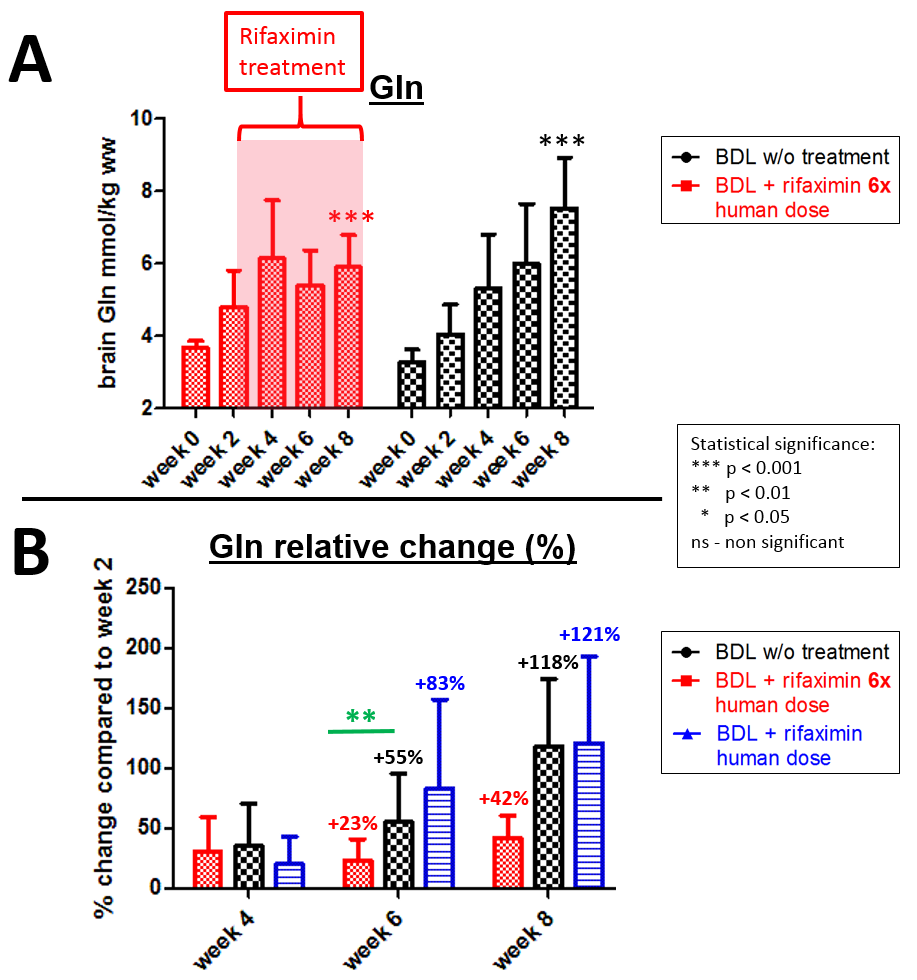
Fig.3
A – Brain Gln evolution from week 0 to week 8 in absolute value in the two groups '6x human-dose rifaximin' BDL rats and BDL rats without treatment.
B – Comparison of brain Gln evolution relative to week 2 in the three groups: BDL rats without treatment, BDL rats treated with human dose, and BDL rats treated with 6x human-dose (‘high-dose’).
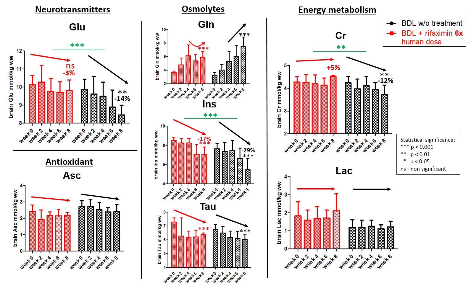
Fig.4 Evolution of some of the main brain osmolytes (Gln, Ins, Tau), metabolites involved in energy metabolism (Lac, Cr), neurotransmitter (Glu) and antioxidant (Asc) in the hippocampus of ‘high-dose rifaximin’ treated group and the group without treatment. Red and black arrows show different trends in the groups.
Two-way ANOVA statistical tests were performed using GraphPad Prism software.