0168
A novel fiber-tracking algorithm using parallel transport frames1Department Neuroscience and Biomedical Engineering, Aalto University, Helsinki, Finland, 2Laboratory of NeuroImaging (LONI), Stevens Neuroimaging and Informatics Institute, University of Southern California, Los Angeles, CA, United States
Synopsis
White matter fiber-tracking algorithms have remarkably improved during the last two decades. However, multiple validation studies warn about the reliability and reproducibility of results, and point out to the need for better algorithms. In propagation based tracking, connections are typically modeled as piece-wise linear segments. In this work, we propose a novel propagation based probabilistic tracker using parallel transport frames which is capable of generating geometrically smooth curves. Moreover, our tracker has a mechanism to reduce noise related propagation errors. Our experiments on FiberCup and Human Connectome Project data show visually and quantitatively superior results compared to three algorithms in MRtrix3.
Introduction
Diffusion MRI (dMRI) based tractography has become an indispensable tool to study the structural connectivity of the brain, in-vivo. Despite significant efforts of the community, tractography has been criticized due to results of validation studies [1,2], importantly with respect to large amounts of false positive [3] and negative connections [4] that are attributed to the complex white matter architecture (fanning, kissing, crossing fibers) of the brain and the noise in the diffusion measurements [5]. These underline the necessity for new tractography algorithms which are capable of addressing the complex geometry of streamlines and at the same time mitigate noise related errors during tracking.
In this work, we present a novel tractography technique that is based on differential geometry of curves and smooth curve parameterizations. In contrast to standard walker based trackers that propagate along straight line segments, the proposed approach takes steps along parametrized smooth curves and is capable of generating highly organized fiber bundles that is useful to study topographic organization of the brain.
Method
The proposed tractography technique operates on fiber orientation density (FOD) images and it is based on parallel transform frames (PTF). PTF is an approach for parameterizing curves using moving frames [6] as follows:$$\begin{bmatrix} \overrightarrow{T}'(t) \\ \overrightarrow{K}'_1(t) \\ \overrightarrow{K}'_2(t) \end{bmatrix}=\begin{bmatrix} 0 & k_1(t) & k_2(t) \\ -k_1(t) & 0 & 0 \\-k_2(t) & 0 & 0\end{bmatrix}\begin{bmatrix} \overrightarrow{T}(t) \\ \overrightarrow{K}_1(t) \\ \overrightarrow{K}_2(t) \end{bmatrix}, \text{for which} ~ \kappa(t) = \sqrt{k_1^2+k_2^2}, $$where $$$\kappa(t)$$$ is curvature and $$$\overrightarrow{T}$$$, $$$\overrightarrow{K}_1$$$ and $$$\overrightarrow{K}_2$$$ are orthonormal vectors akin to the tangent, normal and binormal vectors of the Frenet-Serret moving frame (FSF). In contrast to the well-known FSF however, PTF does not suffer from degeneracy in the case of the vanishing second derivative (straight line) and has advantages for fiber tracking.
Propagation is done similar to the FSF based technique proposed in [7,8]. Instead of a sphere however, we use a probe in our technique, that models a candidate curve segment as a tube enveloping topographically organized curves which is defined by user specified radius and length. The lack of degeneracy in PTF, results with a significantly simpler tracker that comes with a large boost in speed and reduced number of parameters.
Results and discussions
Experimental setup
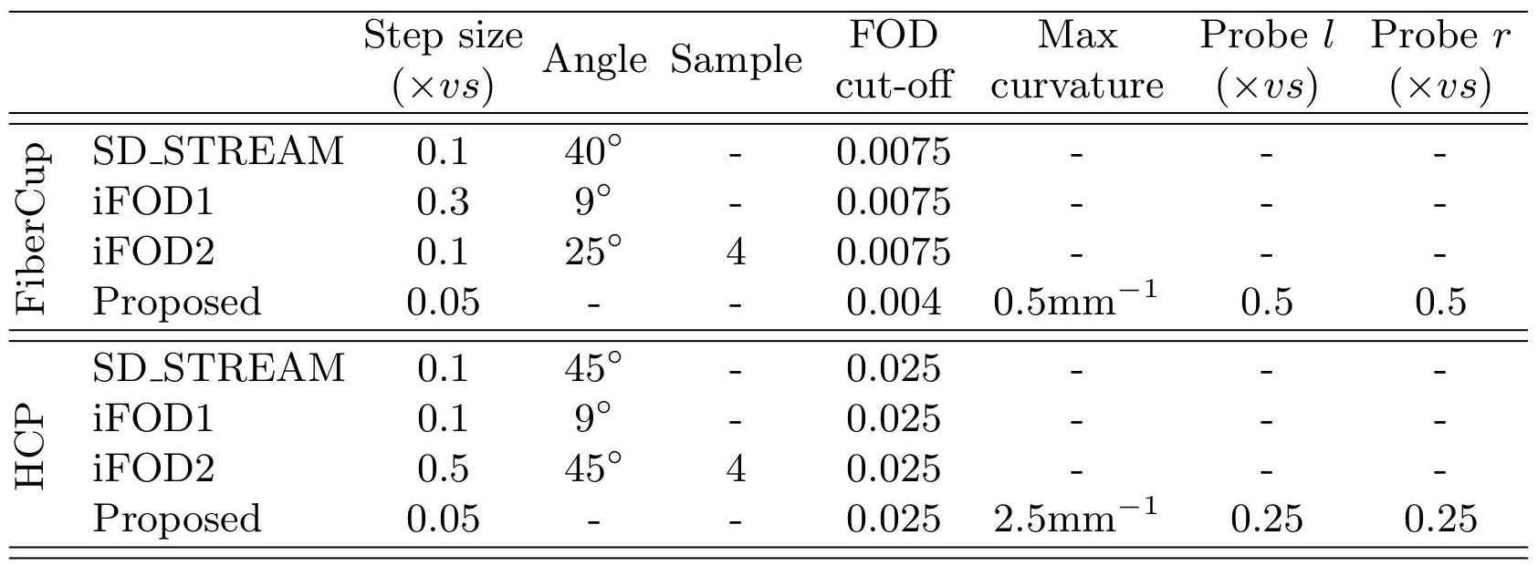
We evaluated our approach both on synthetic and in-vivo datasets against a deterministic (SD_STREAM) and two different probabilistic tractography algorithms (iFOD1, iFOD2) using MRtrix3 (www.mrtrix.org). We used the original FiberCup challenge data since it has ground truth. For in-vivo evaluations, HCP data is used. For all algorithms we used the same FODs computed by the technique proposed in [9]. Tractography parameters are shown in Figure 1.
FiberCup results
For FiberCup tests, we used the average data with b-value 1500s/mm$$$^2$$$. For all techniques, we used the white matter mask as seed image and mask. 100 thousand streamlines were computed with each technique where no post processing nor short track removal were done. With this setup, our results are comparable with those published in the Tractometer study [10].
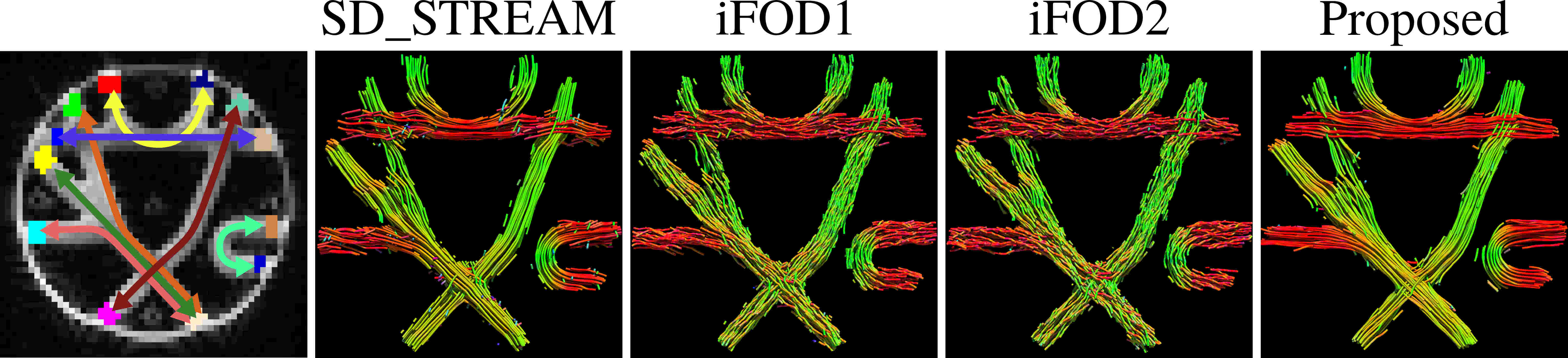
Figure 2 shows visual comparison of the results. Probabilistic iFOD1 and iFOD2 have difficulties propagating along straight lines and lose organization. SD_STREAM and the proposed technique however are visually well organized.
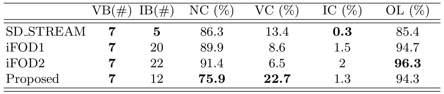
Figure 3 shows results obtained using the Tractometer protocol. The proposed approach gives noticeably good scores for valid connection ratio at the same time yields the least streamlines that do not connect two regions. The proposed approach offers advantages of both the deterministic SD_STREAM technique for yielding low numbers of invalid bundles/connections and the probabilistic iFOD1, iFOD2 techniques for achieving high overlap with ground truth.
HCP results
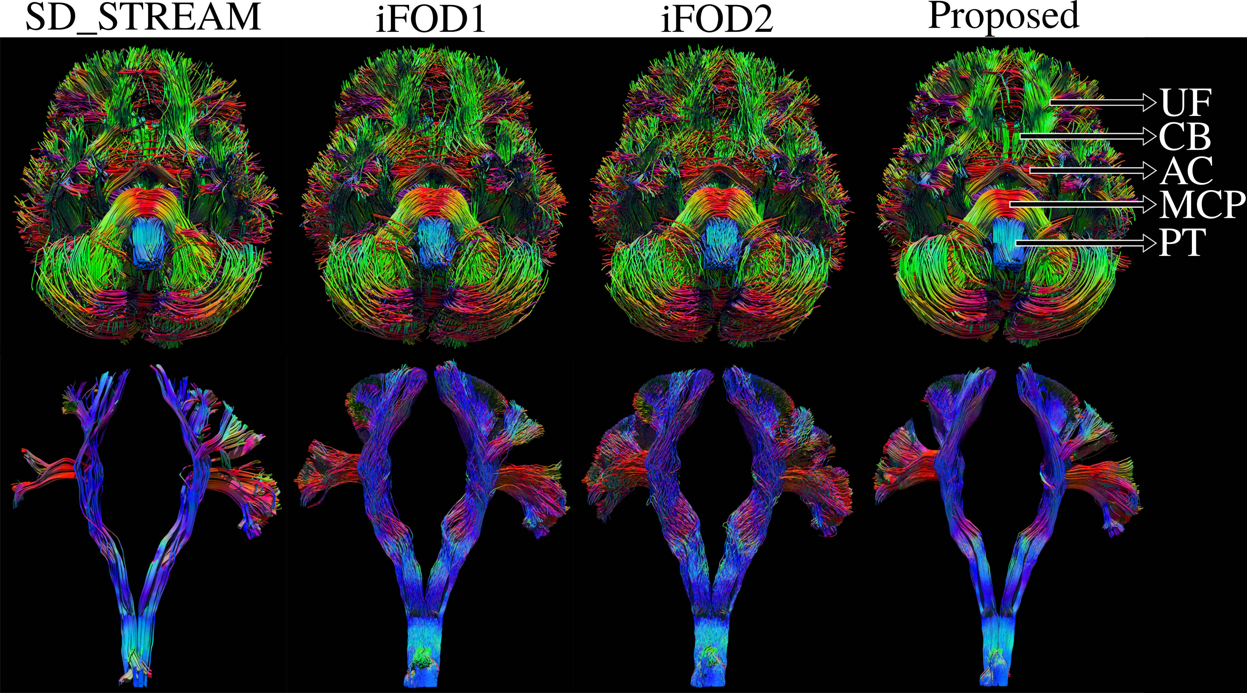
We used the HCP subject 100307 to visually compare whole brain tractograms and pyramidal tracts (PT). For whole brain, we reconstructed 10 thousand streamlines longer than 10cm to highlight long range connections. For PT we computed 6 thousand streamlines where dangling ones were removed using a custom written MATLAB code.
Figure 4 shows whole brains from the inferior view. We observe that the proposed approach produces highly organized bundles where major connections are easily discernible in contrast to iFOD1 and iFOD2. Similarly, SD_STREAM has very organized connections however it fails to capture important cortical projections.
Conclusions
In this work, we proposed a novel tractography algorithm that is capable of generating geometrically smooth curves. At the same time our algorithm takes advantage of topographic regularity to counter noise during tracking. Our implementation runs at affordable speeds, capable of generating ~1K streamlines/minute for typical whole brain tractography using a 4-core (2.4GHz) Intel Core-i7 CPU. The proposed algorithm has intriguing properties with highly promising results that both visually and quantitatively show superior performance compared to state-of-the-art techniques.Acknowledgements
This work was in part supported by the National Institute of Health (NIH) under grants RF1AG056573 and R01EB022744 .References
[1] Thomas C, Ye FQ, Irfanoglu MO, Modi P, Saleem KS, Leopold DA, et al. Anatomical accuracy of brain connections derived from diffusion MRI tractography is inherently limited. PNAS. 2014 Nov 18;111(46):16574–9.
[2] Schilling KG, Nath V, Hansen C, Parvathaneni P, Blaber J, Gao Y, et al. Limits to anatomical accuracy of diffusion tractography using modern approaches. NeuroImage. 2019 Jan 15;185:1–11.
[3] Maier-Hein KH, Neher PF, Houde J-C, Côté M-A, Garyfallidis E, Zhong J, et al. The challenge of mapping the human connectome based on diffusion tractography. Nature Communications. 2017 Nov 7;8(1):1349.
[4] Aydogan DB, Jacobs R, Dulawa S, Thompson SL, Francois MC, Toga AW, et al. When tractography meets tracer injections: a systematic study of trends and variation sources of diffusion-based connectivity. Brain Struct Funct. 2018 Jul 1;223(6):2841–58.
[5] Jones DK, Knösche TR, Turner R. White matter integrity, fiber count, and other fallacies: The do’s and don’ts of diffusion MRI. NeuroImage. 2013 Jun;73:239–54.
[6] Bishop RL. There is More than One Way to Frame a Curve. The American Mathematical Monthly. 1975; 82(3)246–251
[7] Aydogan DB, Shi Y. Probabilistic Tractography for Topographically Organized Connectomes. MICCAI 2016. p. 201–209.
[8] Aydogan DB, Shi Y. Tracking and validation techniques for topographically organized tractography. NeuroImage. 2018 Nov 1;181:64–84.
[9] Tran G, Shi Y. Fiber Orientation and Compartment Parameter Estimation From Multi-Shell Diffusion Imaging. IEEE Transactions on Medical Imaging. 2015 Nov;34(11):2320–32.
[10] Côté M-A, Girard G, Boré A, Garyfallidis E, Houde J-C, Descoteaux M. Tractometer: Towards validation of tractography pipelines. Medical Image Analysis. 2013 Oct;17(7):844–57.
Figures