0160
Can Intravoxel Incoherent Motion Diffusion-weighted Imaging Be used for Preoperative Assessment of Microvascular Invasion in Hepatocellular Carcinoma ?1West China Hospital, Sichuan University, Chengdu, China, 2GE Healthcare China, MR Research, Beijing, China
Synopsis
Microvascular invasion (MVI) is one of the most important factors for the recurrence of hepatocellular carcinoma (HCC), however, accurate preoperative evaluation of MVI is quietly difficult because of the controversy results caused by the conventional imaging features. Compared with diffusion-weighted imaging (DWI), Intravoxel incoherent motion (IVIM) diffusion-weighted MR imaging could better characterize heterogeneity and irregularity of tissue components, and thus may have the potential to better evaluate MVI. In this study, we prospectively determine the usefulness of IVIM parameters and conventional radiologic features for preoperative prediction of MVI in patients with HCC.
Synopsis
Microvascular invasion (MVI) is one of the most important factors for the recurrence of hepatocellular carcinoma (HCC), however, accurate preoperative evaluation of MVI is quietly difficult because of the controversy results caused by the conventional imaging features. Compared with diffusion-weighted imaging (DWI), Intravoxel incoherent motion (IVIM) diffusion-weighted MR imaging could better characterize heterogeneity and irregularity of tissue components, and thus may have the potential to better evaluate MVI. In this study, we prospectively determine the usefulness of IVIM parameters and conventional radiologic features for preoperative prediction of MVI in patients with HCC.Purpose
To prospectively evaluate the potential role of IVIM and conventional radiologic features for preoperative prediction of MVI in patients with HCC.Materials and Methods
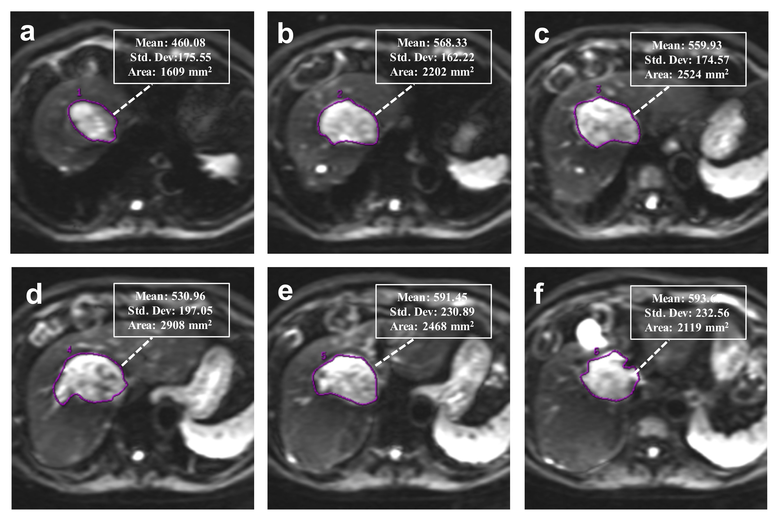
Institutional review board approval and written informed consent were obtained for this study.A cohort comprising 115 patients with 135 newly diagnosed HCCs between January 2016 and April 2017 were evaluated. For all examinations, studies were carried out by using a 3.0 T MR system (Discovery MR 750, GE Healthcare, Milwaukee, USA). A sixteen-channel phased-array torsor coil (GE Medical System) was used for all measurements. IVIM was performed by using an echo-planner imaging sequence with respiratory gating, the parallel imaging was used to short the scanning time and reduce image distortion. The IVIM-DW MR imaging was performed before the injection of contrast agents. Twelve b values from 0 to 1000 sec/mm2 (0, 10, 20, 40, 80, 100, 150, 200, 400, 600, 800 and 1000 sec/mm2) were obtained, and the number of excitations (NEX) for each b value was 1, 6, 4, 2, 2, 2, 1, 1, 2, 4, 6 and 6, respectively. All the IVIM-DW images were analyzed by two independent radiologists blindly, the whole tumor volume was selected for the region of interest (ROI) measurement (Figure 1). Each radiologist drew freehand ROI to outline the tumor on the original DW images (b=400) on each tumor slice, and try to avoid the hemorrhage, calcified and necrotic areas. The ADC, ADCslow, ADCfastand f value were automatically calculated by the workstation, and the averaged value of all tumor slices of each parameter was used for further statistical analyze. Interobserver agreement were checked, univariate and multivariate logistic regression were used for screening the risk factors. Receiver operating characteristics (ROC) curves analyses were performed to evaluate the diagnostic performance.Results
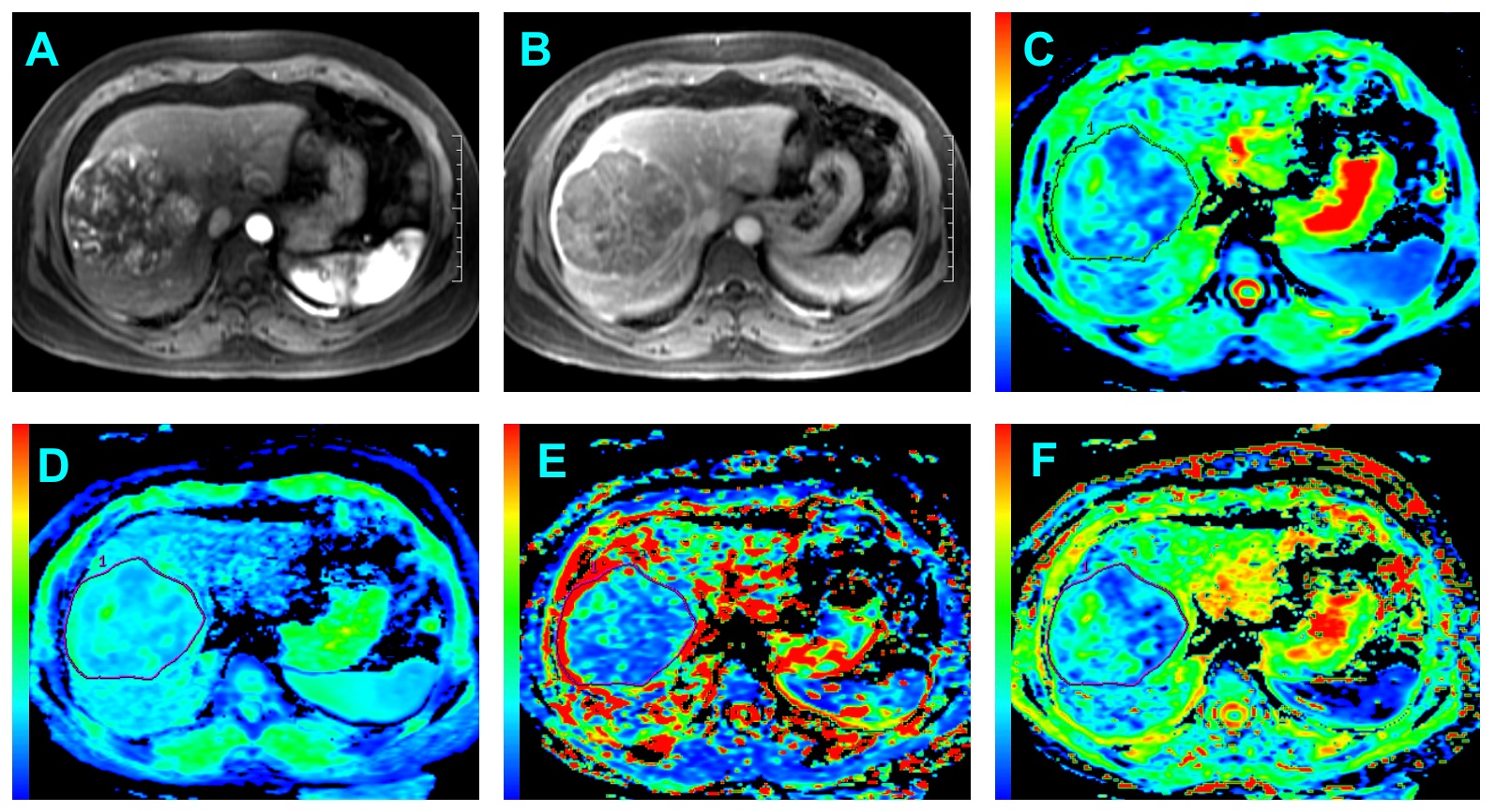
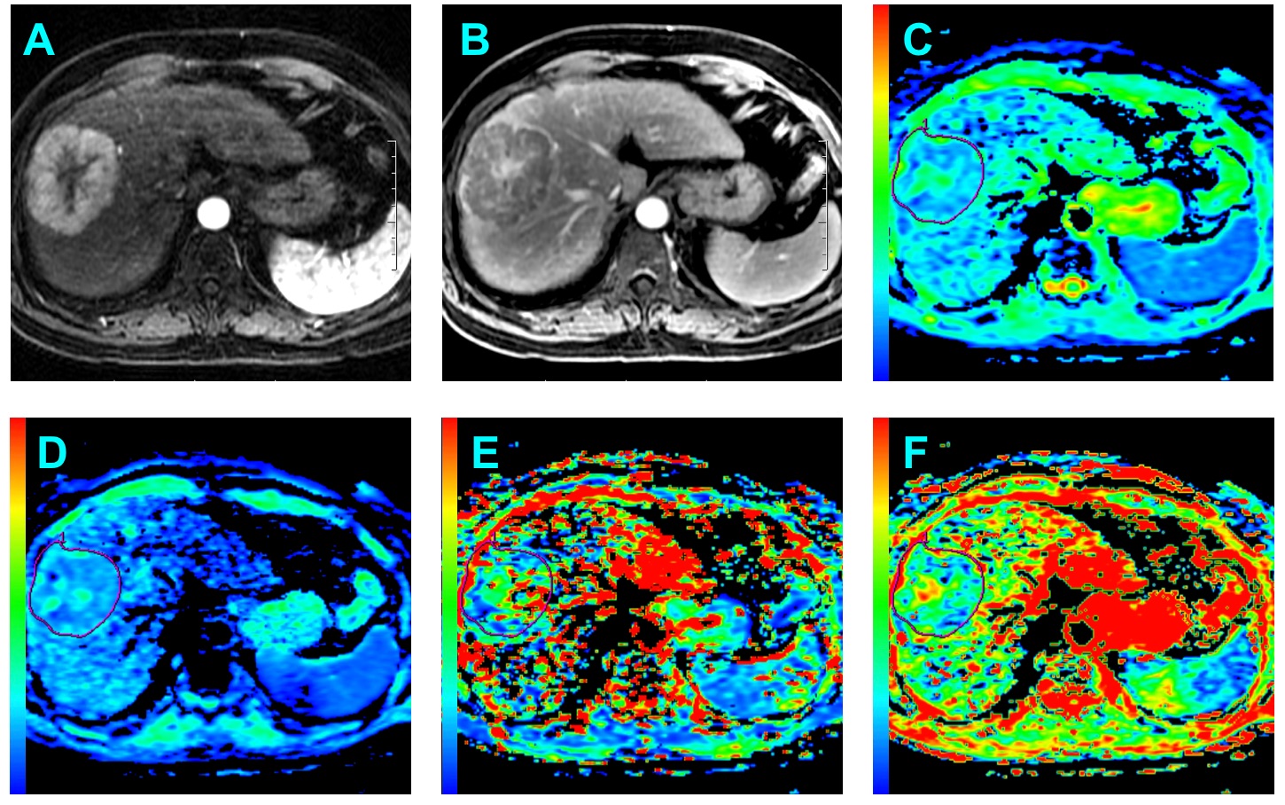
: Good to perfect interobserver agreement was found between two observers regarding the imaging features (all Kappa>0.7). The ADC value measured by both two radiologists were significant higher in the MVI-Negative group than the MVI-Positive group (All p<0.001). In addition, the ADCslow value were also significant higher in the MVI-Negative group compared with the MVI-Positive. No statistical significance was obtained from the ADCfast(R1: p=0.103; R2: p=0.093) and f(R1: p=0.745; R2: p=0.724) in those patients with MVI-Positive (Figure 2)compared with MVI-Negative (Figure 3). Features significantly related to MVI of HCC at univariate analysis were reduced ADC (odds ratio, 0.341; p<0.001), ADCslow(odds ratio, 0.141; p<0.001) and irregular circumferential enhancement (odds ratio, 9.908; p<0.001). At multivariate analysis, only ADCslow(odds ratio, 0.096; p<0.001) was the independent risk factor for MVI of HCC. The mean ADCslow value for MVI of HCC showed an area under ROC curves of 0.815 (95% CI: 0.740-0.877).Conclusion and Discussion
In the present study, the difference of diffusion parameters and the radiologic features were evaluated between the MVI-Positive and MVI-Negative groups and all the risk factors were further screened by using univariate and multivariate logistic regression analysis. The results demonstrated that the decreased ADC and ADCslow value were significantly with the presence of MVI in HCC at univariate analysis. Furthermore, the multivariate analysis suggested that only the ADCslowbased on the IVIM model was the risk factor for MVI and which yield better diagnostic performance in comparison with ADC derived from the mono-exponential model. Thus, the results of the preliminary study have demonstrated that the decreased ADCslowvalue was independent risk factor for predicting MVI of HCC.Acknowledgements
No acknowledgement found.References
1.Torre LA, Bray F, Siegel RL, Ferlay J, Lortet-Tieulent J, Jemal A (2015) Global cancer statistics, 2012. CA Cancer J Clin 65:87-108.
2.Kudo M, Trevisani F, Abou-Alfa GK, Rimassa L (2016) Hepatocellular Carcinoma: Therapeutic Guidelines and Medical Treatment. Liver Cancer 6:16-26.
3. Forner A, Llovet JM, Bruix J (2012) Hepatocellular carcinoma. Lancet 379:1245-1255.
4. Hirokawa F, Hayashi M, Asakuma M, Shimizu T, Inoue Y, Uchiyama K (2016) Risk factors and patterns of early recurrence after curative hepatectomy for hepatocellular carcinoma. Surg Oncol 25:24-29.
5. Rodriguez-Peralvarez M, Luong TV, Andreana L, Meyer T, Dhillon AP, Burroughs AK (2013) A systematic review of microvascular invasion in hepatocellular carcinoma: diagnostic and prognostic variability. Ann Surg Oncol 20:325-339.
Figures