0138
Texture analysis based on diffusional kurtosis imaging for the differentiation of benign and malignant bone tumors1First affiliated hospital of Zhengzhou university, Zhengzhou, China, 2Clinical Science, Philips Healthcare, Beijing, China
Synopsis
This work investigated and evaluated the role of textures extracted from magnetic resonance (MR) diffusion kurtosis imaging (DKI) in characterizing the bone tumors, and furtherly evaluate the ability of these textures to differentiate benign and malignant tumors by using support vector machine classifiers (SVM), which might be helpful for clinical diagnosis and studies. The texture parameters have the ability to character the bone tumor and SVM classifier showed good performance in the differentiation of benign and malignant bone tumors.
Purpose
Recent studies have proved DKI as a reliable imaging technique to evaluate the non-gaussian diffusion behavior in complex biological tissues1,2. And the distribution of the DKI-derived parameters within the tumors might have the potential to describe the characteristics of the whole lesions. In this work, the 3D whole volume texture analysis based on DKI-derived parameter maps were carried out to investigate the application on bone tumors and furtherly evaluate the ability of these texture parameters to differentiate benign and malignant tumors by using support vector machine (SVM) classifier.Methods
Thirty-five patients (20 males and 15 females aged 35.1±19.6 years old) with bone tumors (17 for benign tumors and 18 for malignancies) were included in this study. All the patients were imaged using DKI protocol based on a 3T MR scanner (Ingenia, Philips Healthcare, Best, the Netherlands). The DKI scanning was carried out with 3 b-values of 0, 600, 1,200 s/mm2 and 15 motion-sensitive gradient directions. Mean diffusivity (MD),fractional anisotropy(FA), axial diffusivity (AD), radial diffusivity (RD), mean kurtosis (MK), axial kurtosis(AK) and radial kurtosis (RK) were calculated by using DKE software (Version 2.6.0, website:www.musc.edu/cbi). The whole volume 3D ROI of the lesion were drawn according to the b0, b600 or b1200 image and then the 3D ROI were copied to the above parameter maps for the following texture calculation. Three dimensional texture calculations were carried out by using a modified radiomics-master Matlab toolbox for each parameter map as well as b0, b600 and b1200 images. And for each parameter, 43 textures (Table 1)were extracted and finally 43×10 textures were obtained. Firstly, for all the 43×10 textures, Two sample student’s T test were performed to find the difference between benign and malignant groups. A P value of less than 0.05 was considered statistically significant. And then the textures were reordered by fisher score which indicated the importance of the features. Finally, SVM classification with the reordered features was performed to identify the malignant from the benign bone tumors.Results
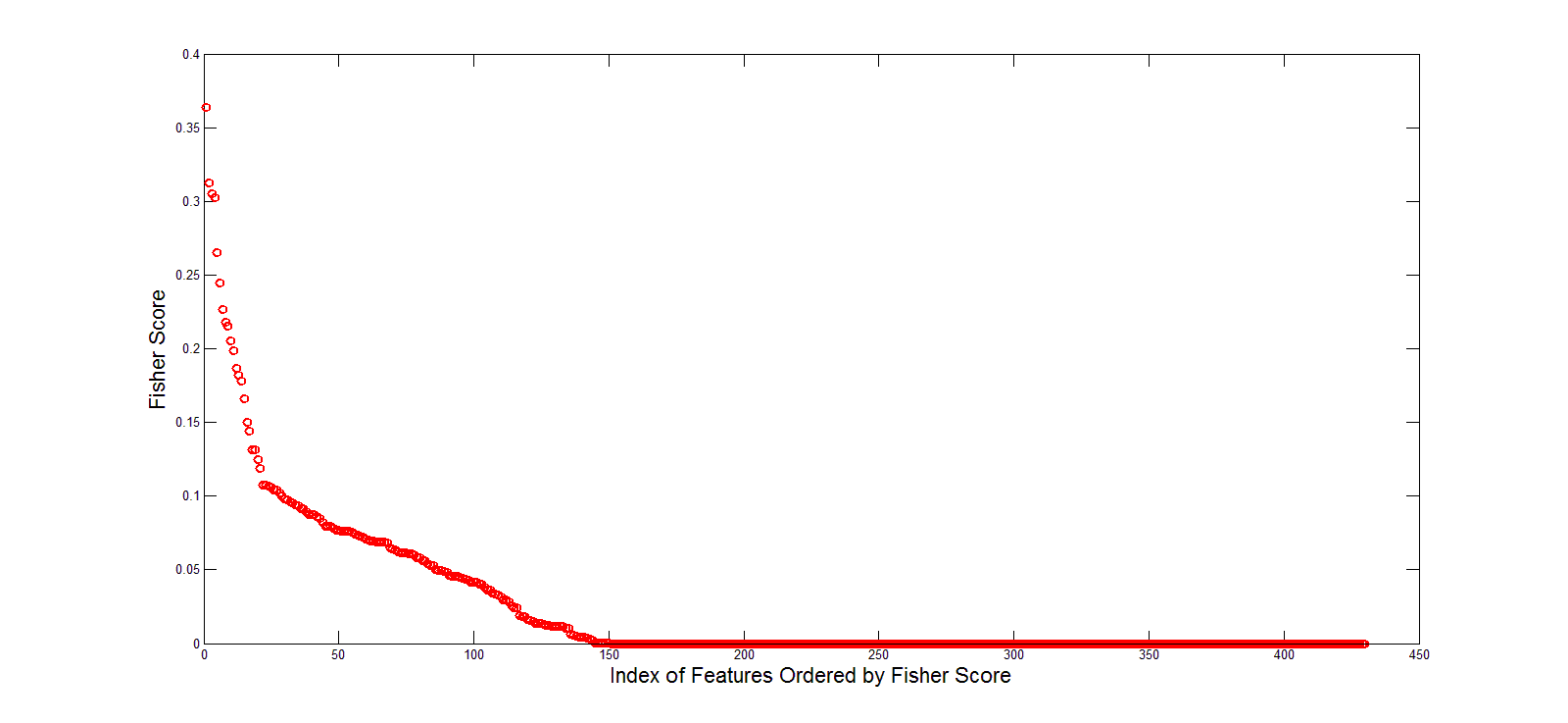
The results revealed that many texture parameters showed a significant difference between benign and malignant bone lesions (P<0.05) and the details were summarized in Figure 1 (because there are too much textures, so we just showed the P value distribution of the features).The reordered features by using fisher score value were shown in Figure 2. The classification results of the benign and malignant bone lesion were summarized in Figure 3. The results showed that the SVM showed a good performance along the features with a high classification accuracy (about 75%-88%), sensitivity (about 78%-91%) and specificity (about 63% -86%).Discussion
DKI is a non-invasive functional imaging based on diffusion MRI technique, which provides useful information of tumor cytoarchitectonic complexityon the water diffusion properties3. The distribution of DKI-derived parameters could reflect the characteristics of the whole lesion and thus might be helpful for the differential diagnosis of benign and malignancy tumors. Different types of tumors have the different microstructure. Therefore, the textures can be applied to reflect the specific microstructure for a specific type of tumors. Our study results show that the texture features extracted from DTI and DKI related parameters is able to differentiate benign from malignant bone tumors. And the SVM classification results showed some texture feature are effective in the differentiation of benign and malignant bone lesion with high accuracy, sensitivity and specificity. This would be very helpful for the clinical diagnosis and therapy evaluation.Conclusions
Texture analysis based on DKI-derived parameters is helpful to evaluate the pathological behavior and provide useful information related to bone tumors microstructure. The multivariate pattern analysis based on textures increases diagnostic confidence of bone tumors.Acknowledgements
No acknowledgement found.References
[1]. Peter Raab, Elke Hattingen, Kea Franz, et al. Cerebral Gliomas: Diffusional Kurtosis Imaging Analysis of Microstructural Differences[J].Radiology,2010,254(3):876-881.
[2]. LuísaNogueira, Sofia Brandão, Eduarda Matos, et al. Application of the diffusion kurtosis model for the study of breast lesions[J].Eur Radiol,2014,24:1197-1203.
[3].Dongmei Wu, Guanwu Li, Junxiang Zhang, et al. Characterization of Breast Tumors Using Diffusion Kurtosis Imaging (DKI)[J].PLOS ONE,2014,9(11):e113240.
Figures