0133
Robust Motion-Compensated Lumbar Spine Bone imaging using 3D UTE with Broadband Inversion Recovery Pulse and k-space Weighted Navigator Gating1Philips Japan, Tokyo, Japan, 2Philips Australia & New Zealand, North Ryde, Australia, 3Qscan Radiology Clinics, Brisbane, Australia, 4Radiology, Eastern Chiba Medical Center, Chiba, Japan, 5General Medical Services, Chiba University Graduate School of Medicine, Chiba, Japan, 6Orthopaedic Surgery, Eastern Chiba Medical Center, Chiba, Japan, 7Philips Healthcare, Tokyo, Japan
Synopsis
We proposed a new technique for the lumbar spine MR bone imaging based on broadband inversion recovery prepared segmented multispoke UTE sequence with k-space weighted navigator gating (3D BoneVIEW) for assessment of low back pain. 3D BoneVIEW provided robust bone imaging with sufficient background suppression and without respiratory artifacts. This sequence has a great potential to help more accurate assessment of the low back pain as an alternative to CT imaging.
Purpose
Low back pain (LBP) is a common disorder involving the muscles, nerves, and bones of the back1. MRI provides valuable information regarding the underlying causes of LBP2. For example, the extraforaminal nerve root entrapment3 can be a cause of low back pain; therefore, it is important to evaluate pathologic lesions in the extraforaminal area. Computed tomography (CT) provides bone information, it helps to assess the relationship between entrapped nerve and surrounding bone structure before and after surgical treatment4.
Recently, MR bone imaging, using ultrashort echo-time (UTE) or zero echo-time (ZTE) sequence, has gained more attention for detection and assessment of bone pathology as an alternative to CT imaging5-8. Challenge of MR bone imaging for clinical application to the lumbar spine is to increase the robustness of image quality. Bone weighted imaging suffers from insufficient background suppression due to B0 inhomogeneities. Furthermore, respiratory motion adversely affects the image quality in the lumbar spine, it results in increasing the image noise and streak artifacts.
To overcome these problems, we proposed a new technique based on broadband inversion recovery (IR) prepared segmented multispoke UTE sequence with k-space weighted navigator gating (3D BoneVIEW). The purpose of this study was to evaluate the feasibility of 3D BoneVIEW in the lumbar spine for assessment of LBP.
Methods
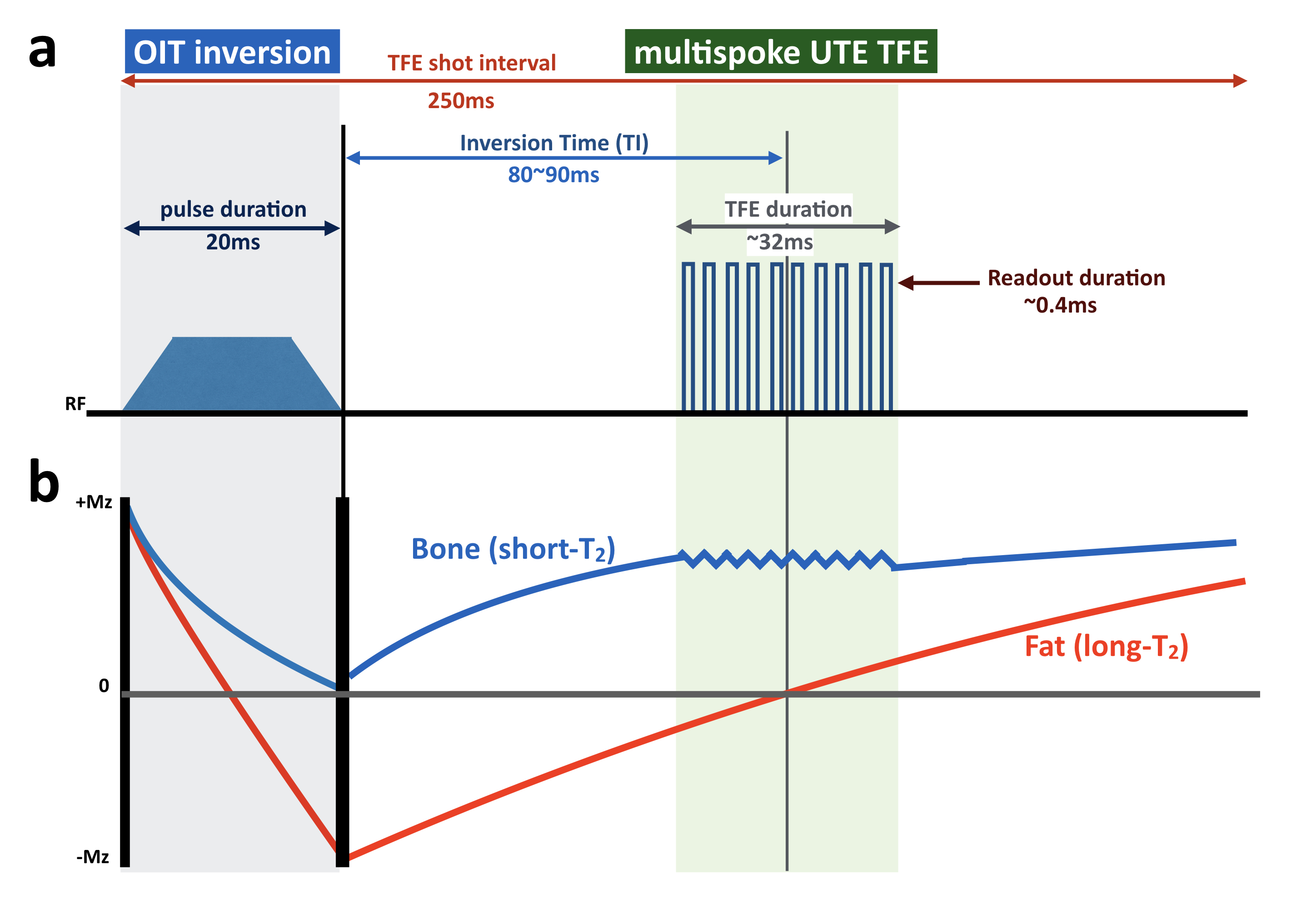
3D BoneVIEW is based on IR-prepared UTE 3D stack-of-stars radial sequence9 (IR-TFE)[Fig.1a]. Adiabatic inversion pulses selectively invert long-T2 species and fat simultaneously10. For robustness against B1 and B0 sensitivity, we applied a broadband offset independent trapezoid (OIT) pulse, which has already been used for brachial plexus MR neurography11,12. To suppress the background signals sufficiently, we used a long duration OIT inversion pulse (≥20ms). By using such a longer pulse duration, long T2 species are inverted whereas short T2 species are saturated9 because the T2 of bone is significantly shorter than the duration of the RF pulse. The TFE shot interval and inversion delay (TI) are chosen such that optimal nulling of the signal from both muscle and fat13 is achieved [Fig.1b]. This provides high contrast morphological imaging of bone.
To prevent the blurring of the bone due to T2* decay during the readout period, literature14 suggests that readout duration should be less than 0.81xT2*. Since the T2 value of cortical bone is on the order 400us15, we kept the actual readout duration within 400us. For the TFE multispoke excitation, a shot duration of 250ms was applied because the T1 of bone is around 250ms. In addition, we set the constant shot length (actual TRxTFE factor) around 32ms to prevent the recovery of fat signals during TFE shot.
Regarding respiratory compensation, k-space weighted gating16-18 was used in this study. Weighted gating is an extension of the respiratory navigator gating. It allows to use a different gate width for the periphery of k-space than for the center of k-space. Using a larger gate for the periphery of k-space will reduce the scan time significantly. The navigator was put on the anterior abdominal wall and applied with A-P direction and captured the abdominal respiratory motion (around the lumbar spine) directly. Consequently, it can clean up the streak artifact; therefore, we can use a lower density of radial angles and reduce scan time further.
A total of ten volunteers and two patients with LBP were examined with 3.0T or 1.5T whole-body clinical system (Ingenia, Philips Healthcare). The study was approved by the local IRB, and written informed consent was obtained from all subjects.
Imaging parameters for BoneVIEW were; 3D stack-of-stars radial UTE-TFE, Coronal acquisition, voxel size=1.4*1.4*3mm, FOV=400*400mm, TFE shot interval=250ms, flip angle=16°, turbo factor=11, TR/TE=2.9/0.09ms, TI=94ms, weighted navigator gating (gating window for central/peripheral k-space=5/10mm), and total acquisition time=7 to 10minutes (depend on the body habitus).
Results and Discussion
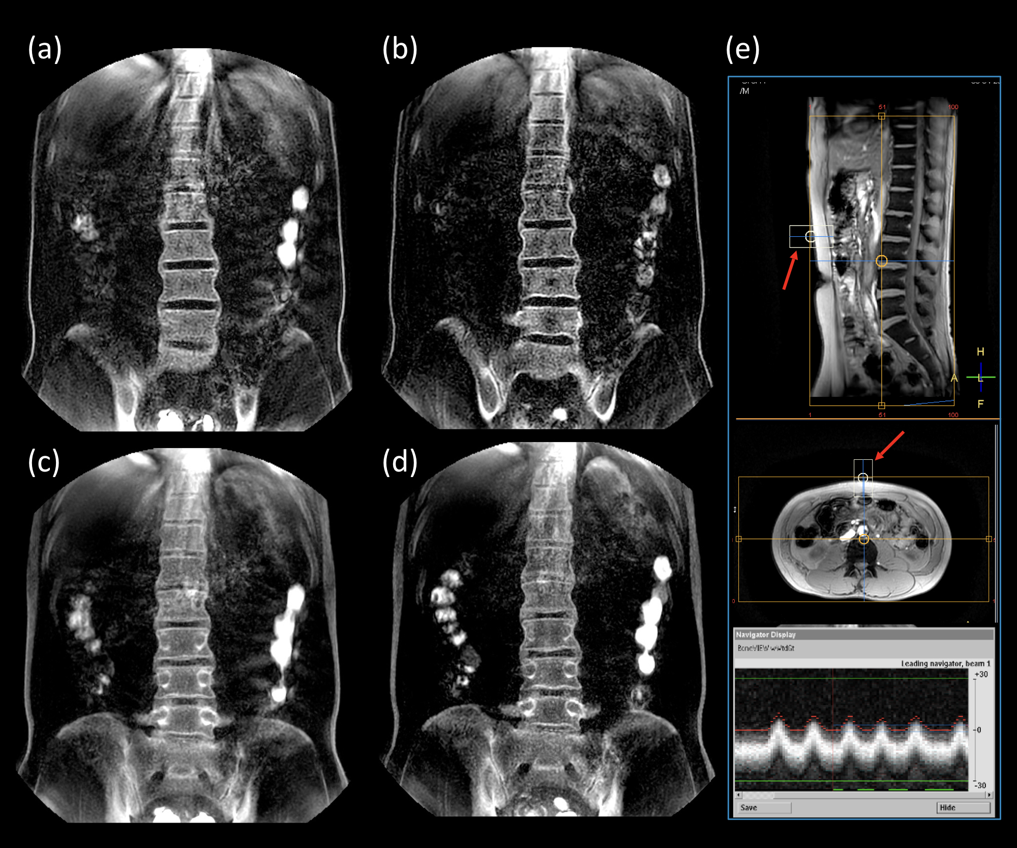
Figure 2 demonstrates the effect of weighted gating. Compared to a non-gated free-breathing scan [Fig.2a], weighted gating reduced the image noise and streak artifacts due to respiratory motion [Fig.2b] without too much prolongation of scan time.
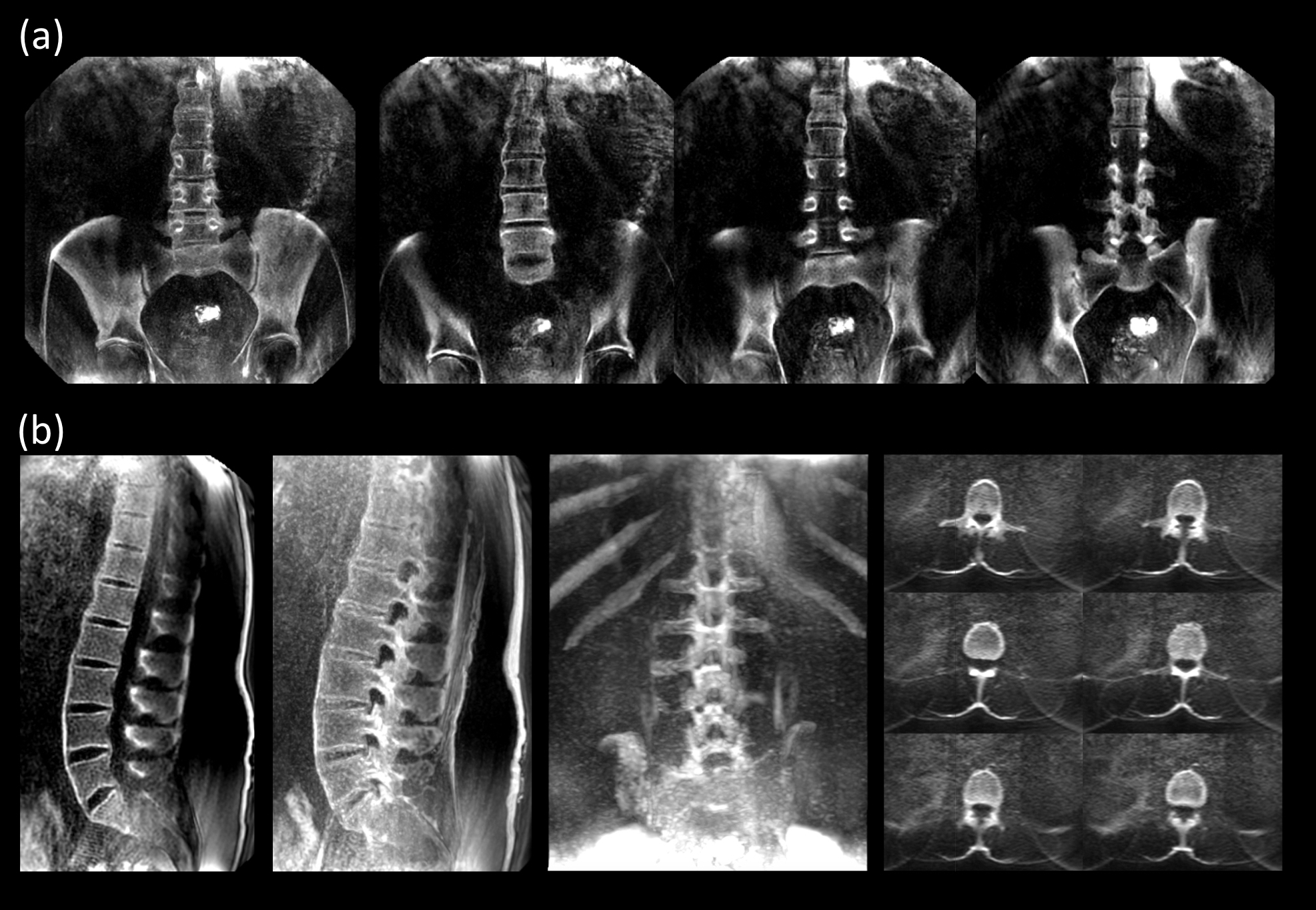
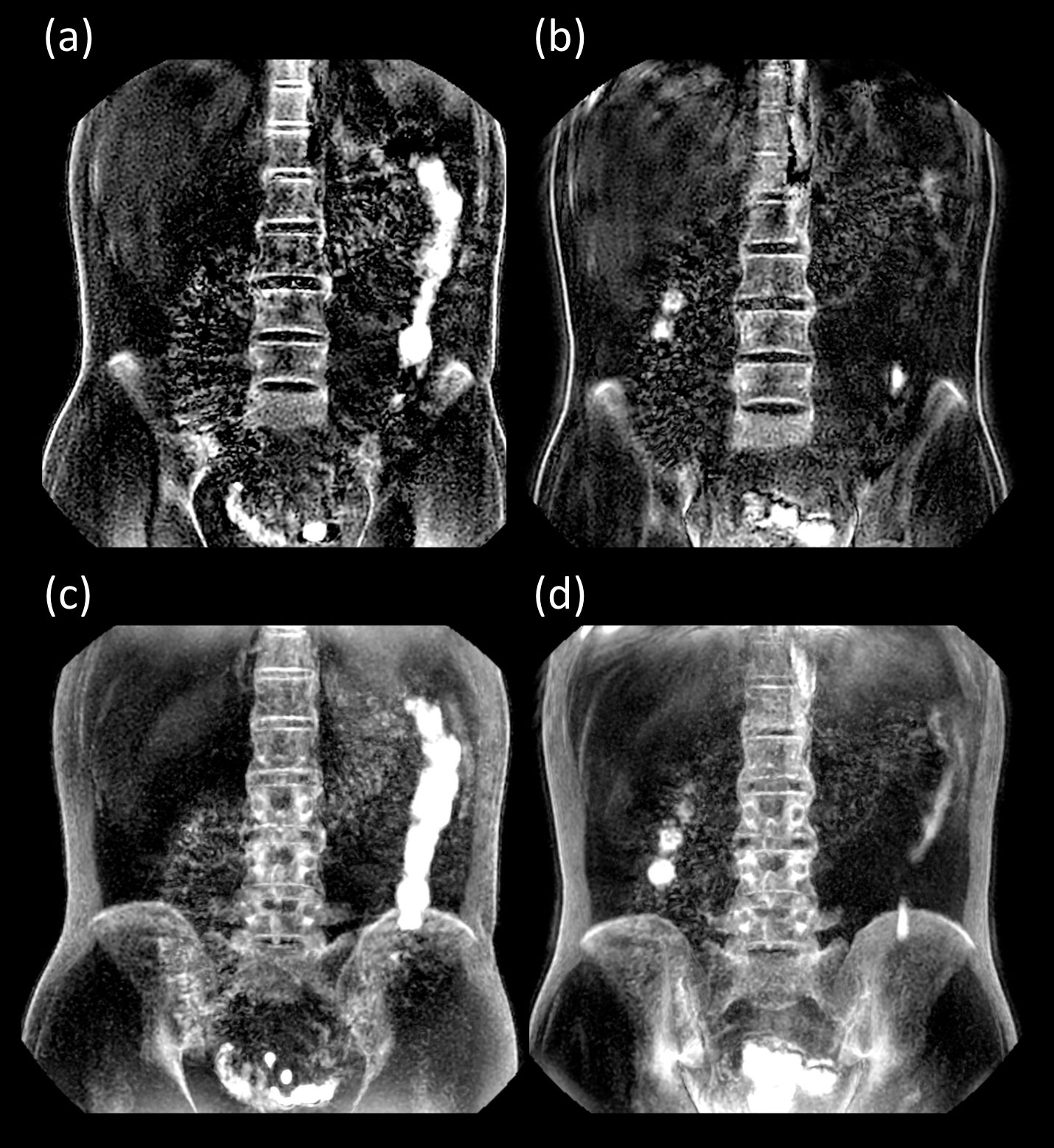
Representative coronal and sagittal BoneVIEW images with partial MIP are shown in Figure 3. BoneVIEW clearly depicted the cortical bone structure of the lumbar spine while suppressing background signals sufficiently. Furthermore, Figure 4 shows the comparison between 3.0T and 1.5T with same volunteer and almost same scan parameters. BoneVIEW provided similar image contrast at both field strength.
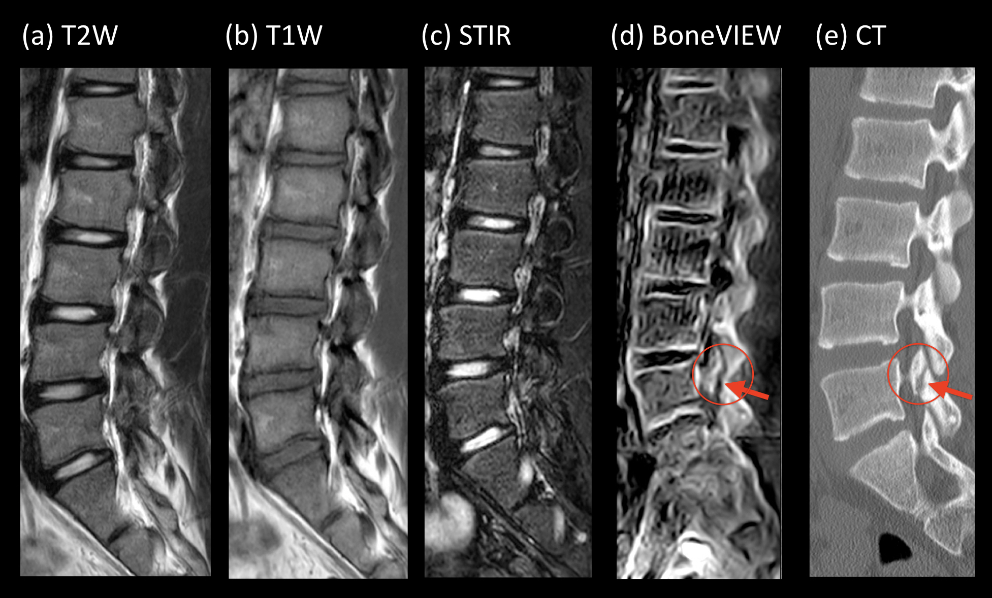
Figure 5 shows representative clinical case in a patient with L5 spondylolysis comparison with CT scan. BoneVIEW provided comparable information with CT imaging.
Conclusion
This study showed a new scheme for MR bone imaging of the lumbar spine by using respiratory motion-compensated IR-UTE sequence. This sequence has a great potential to help more accurate assessment of the LBP as an alternative to CT imaging.Acknowledgements
No acknowledgement found.References
1. ”Low Back Pain Fact Sheet". National Institute of Neurological Disorders and Stroke. 3 November 2015. https://www.ninds.nih.gov/Disorders/Patient-Caregiver-Education/Fact-Sheets/Low-Back-Pain-Fact-Sheet
2. Roudsari B, et al. Lumbar spine MRI for low back pain: indications and yield. AJR Am J Roentgenol. 2010;195:550-9.
3. Heo DH, et al. Simple oblique lumbar magnetic resonance imaging technique and its diagnostic value for extraforaminal disc herniation. Spine 2009;34:2419–23.
4. Eguchi Y, et al. Discrimination between Lumbar Intraspinal Stenosis and Foraminal Stenosis using Diffusion Tensor Imaging Parameters: Preliminary Results. Asian Spine J. 2016;10:327-34.
5. Delso G, et al. Clinical evaluation of zero-echo-time MR imaging for the segmentation of the skull. J Nucl Med. 2015;56:417-22.
6. Wiesinger F, et al. Zero TE MR bone imaging in the head. Magn Reson Med. 2016;75:107-14.
7. Nazaran A, et al. Three-dimensional adiabatic inversion recovery prepared ultrashort echo time cones (3D IR-UTE-Cones) imaging of cortical bone in the hip. Magn Reson Imaging. 2017;44:60-64.
8. Wiesinger F, et al. Zero TE-based pseudo-CT image conversion in the head and its application in PET/MR attenuation correction and MR-guided radiation therapy planning. Magn Reson Med. 2018;80:1440-1451.
9. Carl M, et al. UTE imaging with simultaneous water and fat signal suppression using a time-efficient multispoke inversion recovery pulse sequence. Magn Reson Med. 2016;76:577-82.
10. Larson PE, et al. Using adiabatic inversion pulses for long-T2 suppression in ultrashort echo time (UTE) imaging. Magn Reson Med. 2007;58:952-61.
11. Yoneyama M, et al. Motion-Sensitized Driven-Inversion (MSDI) for improvement of diffusion-prepared MR neurography (SHINKEI) in the brachial plexus. Proc Intl Soc Mag Reson Med. 2017;25:0854.
12. Yoneyama M, et al. Quantitative MR Neurography with Robust Fat Suppression. Proc Intl Soc Mag Reson Med. 2018;26:5400.
13. Li S, et al. Effects of inversion time on inversion recovery prepared ultrashort echo time (IR-UTE) imaging of bound and pore water in cortical bone. NMR Biomed. 2015;28:70-8.
14. Gai ND, et al. Optimized Ultra-short Echo Time Breathhold 3D Lung Imaging. Proc. Intl. Soc. Mag. Reson. Med. 2015;23:3977.
15. Carl M, et al. Radiofrequency pulses for simultaneous short T2 excitation and long T2 suppression. Magn Reson Med. 2011;65:531-7.
16. Muthupillai R, et al. SENSE or k-MAG to accelerate free breathing navigator-guided coronary MR angiography. AJR Am J Roentgenol 2006;186:1669-1675.
17. Akcakaya M, et al. Free-breathing phase contrast MRI with near 100% respiratory navigator efficiency using k-space-dependent respiratory gating. Magn Reson Med 2014;71:2172-2179.
18. Dyverfeldt P, et al. Comparison of respiratory motion suppression techniques for 4D flow MRI. Magn Reson Med. 2017;78:1877-1882.
Figures