0120
Test-retest Reliability of Myelin-Sensitive MRI Techniques1Health Sciences Progam, University of Calgary, Calgary, AB, Canada, 2Alberta Children's Hospital Research Institute, University of Calgary, Calgary, AB, Canada, 3Biomedical Engineering Graduate Program, University of Calgary, Calgary, AB, Canada, 4Department of Radiology, University of Calgary, Calgary, AB, Canada, 5GE Healthcare, Calgary, AB, Canada
Synopsis
This project evaluated the reliability of quantitative inhomogeneous magnetization transfer (qihMT), a novel myelin-sensitive measure, compared to more common measures, magnetization transfer ratio (MTR) and fractional anisotropy (FA), in 10 healthy adults. A repeated measures ANOVA revealed no significant differences in measure means between scans in 18 white matter regions. Coefficients of variation (CV) demonstrated that FA had the highest reliability, followed by MTR and qihMT. The reduced reliability of qihMT observed here may be mitigated by further optimization of this novel sequence.
Purpose
In vivo assessment of white matter and myelin is important for numerous clinical and research applications.1 Diffusion tensor imaging (DTI) and magnetization transfer (MT) imaging are sensitive and commonly used techniques to assess white matter structure.2,3 Newer techniques, such as inhomogeneous magnetization transfer (ihMT) are becoming more common in research, as they are more specific to myelin.4,5 ihMT provides typical MT measures, including MTR, alongside measures with high myelin sensitivity and specificity, such as quantitative ihMT (qihMT). However, no previous studies have examined the test-retest reliability of ihMT. This project compared the reliability of qihMT to traditional myelin sensitive measures, FA and MTR.Methods
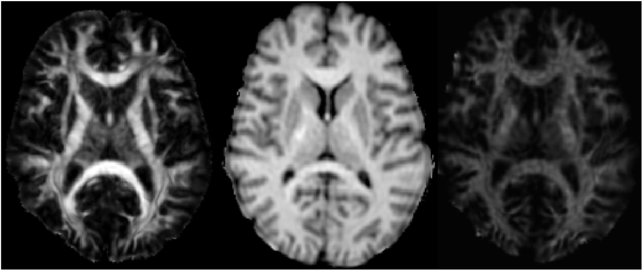
10 healthy volunteers (7 F/3M) were scanned on a 3T GE Discovery MR750w scanner using a 32 channel head coil. DTI and ihMT sequences were each performed twice on two separate days, for a total of four datasets per sequence per participant. DTI: spin echo EPI, TR/TE = 12s/88ms, 2.2mm isotropic resolution, 30 diffusion directions at b = 900 s/mm2, 5 interleaved volumes at b=0 s/mm2, scan time = 7:12. IhMT: 3D SPGR sequence: TR/TE = 10.46ms/2.18ms, 2.2mm isotropic resolution, 8° flip angle, scan time = 5:12. IhMT included a 5ms Fermi pulse with peak B1 of 45 mG and ±5kHz offset prior to each excitation. A 32° flip angle reference image with no MT pulse was acquired for qihMT production.5 FA, MTR, and qihMT maps (Fig. 1) were produced and processed for brain extraction, then registered using the Advanced Normalization Tools (ANTs) toolbox to the JHU-ICBM atlas 1mm FA map.6 Mean FA, MTR and qihMT values were extracted from 18 major regions of the JHU atlas. A repeated measures ANOVA was conducted to compare mean FA, MTR, and qihMT between scan repetitions in each of the 18 white matter regions. Mean coefficient of variations (CV) were calculated for each FA, MTR, and qihMT to evaluate measure stability between scans in 18 white matter regions. A paired t-test was conducted to compare all FA, MTR and qihMT CVs.Results
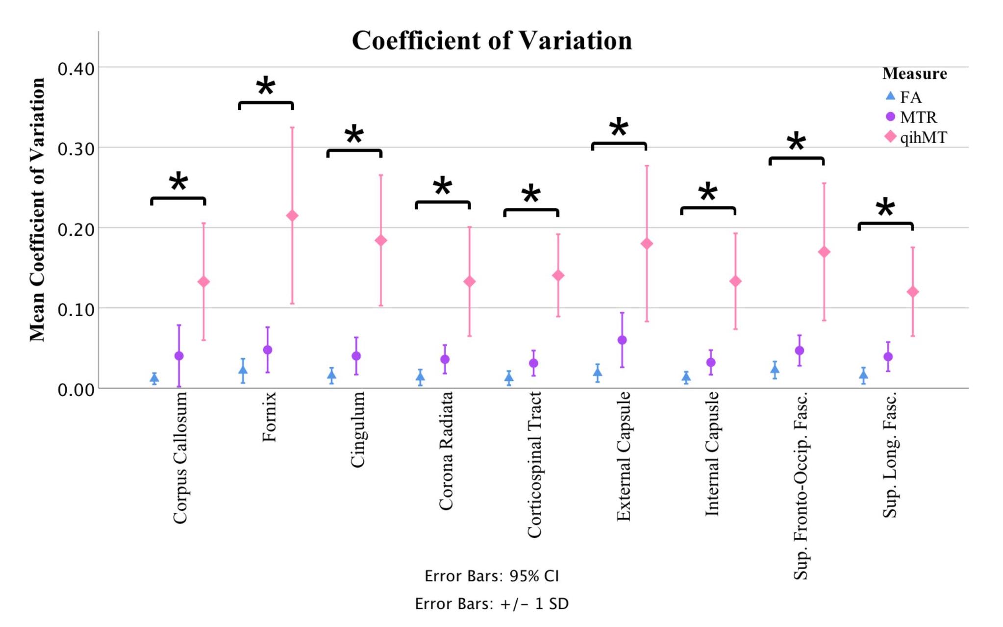
Repeated measures ANOVAs revealed no significant scan-to-scan differences for FA, MTR and qihMT in any examined region. FA had the lowest CV values across all 18 regions, followed by MTR and finally qihMT (Fig. 2); differences between measure CVs were significant. For FA, the average CVs ranged from a low of 0.011 in the right corticospinal tract to a max of 0.025 in the right superior fronto-occipital fasciculus. MTR had a minimum CV of 0.031 in the corpus callosum, and a maximum CV of 0.071 in the right uncinate fasciculus. The CVs for qihMT ranged from 0.109 in the right superior longitudinal fasciculus to 0.311 in the left uncinate fasciculus.Discussion
FA, MTR, and qihMT showed consistent trends of reliability in our analysis, with FA being the most reliable, followed by MTR and qihMT. The lack of significant differences between scans indicates good reliability for each measure within major white matter regions. Quantitatively, FA exhibited consistently low CVs across regions and therefore demonstrated the highest reliability, while more variability was observed in MTR and qihMT. CVs <0.05 are considered to have high reliability, showing that both FA and MTR are highly reliable. qihMT in our study was less reliable, though still showed consistency across scans in the ANOVA. QihMT’s higher CVs may have been driven by ihMT’s lower signal-to-noise ratio, which could be improved by recently proposed optimizations to the technique, including concentrated MT preparation.7 Given the increased specificity of qihMT to myelin, these modifications are worth exploring further.5 Overall, FA, MTR and qihMT demonstrated good reliability in healthy volunteers. Although qihMT demonstrated the lowest reliability (highest CV values) among the three measures, it performed adequately in repeated measures ANOVA analysis, and has been shown to be tightly correlated to other measures of myelin.5Conclusion
QihMT, FA and MTR all demonstrate good reliability in major white matter regions of healthy adult volunteers. This study’s findings inform which myelin-sensitive MRI techniques are used in future studies and clinical practice. Given that qihMT is more specific to myelin than FA and MTR, it may offer insights into the changes that occur in myelin over time, assist physicians in assessing the progression of myelin associated diseases and help evaluate a patient’s response to treatment.Acknowledgements
No acknowledgement found.References
1. Fields RD. White matter in learning, cognition and psychiatric disorders. Trends in Neurosciences. 2008;31(7):361–70.
2. Beaulieu C. The basis of anisotropic water diffusion in the nervous system - a technical review. NMR in Biomedicine. 2002 Dec;15(7-8):435–55.
3. Henkelman RM, Stanisz GJ, Graham SJ. Magnetization transfer in MRI: a review. NMR in Biomedicine. 2001 Apr;14(2):57–64.
4. Varma G, Girard O, Prevost V, Grant A, Duhamel G, Alsop D. Interpretation of magnetization transfer from inhomogeneously broadened lines (ihMT) in tissues as a dipolar order effect within motion restricted molecules. Journal of Magnetic Resonance. 2015 Nov; 260:67–76.
5. Geeraert BL, Lebel RM, Mah AC, Deoni SC, Alsop DC, Varma G, et al. A comparison of inhomogeneous magnetization transfer, myelin volume fraction, and diffusion tensor imaging measures in healthy children. NeuroImage. 2017 Sept.
6. Avants BB, Tustison NJ, Song G, Cook PA, Klein A, Gee JC. A reproducible evaluation of ANTs similarity metric performance in brain image registration. NeuroImage. 2011Feb;54(3):2033–44.
7. Mchinda S, Varma G, Prevost VH, Troter AL, Rapacchi S, Guye M, et al. Whole brain inhomogeneous magnetization transfer (ihMT) imaging: Sensitivity enhancement within a steady-state gradient echo sequence. Magnetic Resonance in Medicine. 2017;79(5):2607–19.
Figures