0115
Reproducibility of Practical Imaging-Based Myelin Biomarkers1Division of Biomedical Engineering, Hankuk University of Foreign Studies, Yongin, Korea, Republic of, 2Imaging Institute, The Cleveland Clinic, Cleveland, OH, United States
Synopsis
Remyelination therapies are an emerging approach for treating multiple sclerosis, but development of these therapies is hampered by a lack of imaging biomarkers. Imaging with improved specificity to myelin, as compared to conventional MRI, have the potential to act as biomarkers, but current implementations can be time consuming. We examine the performance of fast version of myelin imaging from the perspective of reproducibility, a necessary prerequisite for use in proof-of-concept clinical trials of remyelinating agents.
Introduction
Remyelination is an emerging strategy for treating multiple
sclerosis. However, evaluation of remyelination therapy in clinical trials is
hindered by a lack of imaging biomarkers. Myelin water imaging (MWI)1 and quantitative
magnetization transfer (qMT)2 are candidate biomarkers for myelin, but can be difficult to implement in patient studies because of long scan times. Fast
versions have been introduced3,4 as well as an alternative
approach for imaging myelin water, visualization of short transverse relaxation
component (ViSTa)5. We examine the
reproducibility of these methods to determine if they are ready for use in
patient studies.Methods
Four healthy subjects underwent an IRB-approved scan-reposition-rescan protocol on a Siemens 3T Prisma with a standard 20-channel head coil (Siemens Medical Solutions, Erlangen). GRASE-based fast MWI (TA=17:35), fast qMT (TA=5:58) and ViSTA (TA=5:49) were acquired at 1.5x1.5x4 mm3 resolution. An MP2RAGE acquisition6 at the same spatial resolution was also acquired (TA=3:58) to provide T1 maps for calculating macromolecular proton fraction for qMT. A multi-exponential fit was performed to estimate myelin water fraction for MWI. White matter regions of interest (ROI) from the JHU white-matter tractography atlas7 were aligned to native space using FSL8. Intraclass correlation coefficients (ICC)9 were calculated in each ROI and in all white matter to quantify reproducibility.Results
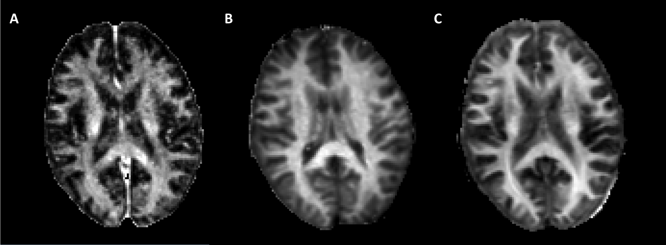
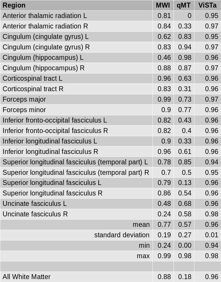
Figure 1 shows myelin density maps for each modality, demonstrating the signal-to-noise-ratio advantage of qMT and ViSTa over MWI. Figure 2 shows ICC across subjects and scans of the mean across all voxels in each of 20 white matter pathways of across all voxels within all the pathways (white matter). Using the guidelines suggested by Cicchetti10, reproducibility can be considered poor for ICC less than 0.40, fair for values from 0.40 to 0.59, good from 0.60 to 0.74 and excellent from 0.75 to 1.00. When considering 20 white matter pathways across the brain, reproducibility ranges from poor to excellent for MWI, poor to excellent for qMT and good to excellent for ViSTa. Averaging across all of white matter, reproducibility is excellent for MWI and ViSTa but poor for qMT.Discussion
While the qualitative signal-to-noise ratio of qMT suggests that it is a good candidate as a biomarker for myelin, the highly variable reproducibility across the brain suggest that the current implementation is not appropriate for use as an imaging biomarker. The overall consistency and excellent reproducibility of ViSTa and suggests that it is the best of the three candidates examined. While none of these imaging methods are direct indicators of myelin density, sensitivity to changes of imaging biomarkers may be sufficient for use in clinical trials of proof-of-concept clinical trial of remyelination11. Future work will focus on the reproducibility of these methods in multiple sclerosis patients to determine the generalizability of these results.Acknowledgements
We thank Siemens Medical Solutions for source code. We thank Tobias Kober and Bénédicte Maréchal of Siemens Medical Solutions for WIP900B.References
1. MacKay, A., Whittall, K., Adler, J., Li, D., Paty, D. & Graeb, D. In vivo visualization of myelin water in brain by magnetic resonance. Magn Reson Med 1994; 31(6):673-677.
2. Sled, J. G. & Pike, G. B. Quantitative interpretation of magnetization transfer in spoiled gradient echo MRI sequences. J Magn Reson 2000; 145(1):24-36.
3. Oh, S. & Lowe, M. in Proceedings 25th Scientific Meeting, International Society for Magnetic Resonance in Medicine. 4738.
4. Oh, S. H., Choi, J. Y., Im, Y., Prasloski, T. & J., L. in Proceedings 23rd Scientific Meeting, International Society for Magnetic Resonance in Medicine. 3143.
5. Oh, S. H., Bilello, M., Schindler, M., Markowitz, C. E., Detre, J. A. & Lee, J. Direct visualization of short transverse relaxation time component (ViSTa). Neuroimage 2013; 83(485-492.
6. Marques, J. P., Kober, T., Krueger, G., van der Zwaag, W., Van de Moortele, P. F. & Gruetter, R. MP2RAGE, a self bias-field corrected sequence for improved segmentation and T1-mapping at high field. Neuroimage 2010; 49(2):1271-1281.
7. Hua, K., Zhang, J., Wakana, S., Jiang, H., Li, X., Reich, D. S., Calabresi, P. A., Pekar, J. J., van Zijl, P. C. & Mori, S. Tract probability maps in stereotaxic spaces: analyses of white matter anatomy and tract-specific quantification. Neuroimage 2008; 39(1):336-347.
8. Smith, S. M., Jenkinson, M., Woolrich, M. W., Beckmann, C. F., Behrens, T. E., Johansen-Berg, H., Bannister, P. R., De Luca, M., Drobnjak, I., Flitney, D. E., Niazy, R. K., Saunders, J., Vickers, J., Zhang, Y., De Stefano, N., Brady, J. M. & Matthews, P. M. Advances in functional and structural MR image analysis and implementation as FSL. Neuroimage 2004; 23 Suppl 1(S208-219.
9. Shrout, P. E. & Fleiss, J. L. Intraclass correlations: uses in assessing rater reliability. Psychol Bull 1979; 86(2):420-428.
10. Cicchetti, D. V. Guidelines, criteria, and rules of thumb for evaluating normed and standardized assessment instruments in psychology. Psychological Assessment 1994; 6(4):284-290.
11. Reich, D. S., White, R., Cortese, I. C., Vuolo, L., Shea, C. D., Collins, T. L. & Petkau, J. Sample-size calculations for short-term proof-of-concept studies of tissue protection and repair in multiple sclerosis lesions via conventional clinical imaging. Multiple sclerosis (Houndmills, Basingstoke, England) 2015; 21(13):1693-1704.
Figures