0112
3D Inversion Recovery Ultrashort Echo Time (3D IR-UTE) Magnetic Resonance Imaging of Myelin in Traumatic Brain Injury – a Feasibility Study1University of California, San Diego, San Diego, CA, United States, 2VA San Diego Healthcare System, San Diego, CA, United States
Synopsis
Reduced myelination has been observed in rodents and humans after mild traumatic brain injury (mTBI). However, conventional MR imaging sequences cannot directly detect any signal from myelin. In this study, we aimed to evaluate a 3D IR-UTE sequence for selective imaging of myelin in mice using a standard controlled cortical impact (CCI) model of mTBI with histological confirmation. To demonstrate the clinical feasibility, a translational 3D IR-UTE technique was developed and applied to healthy volunteers and mTBI patients at 3T. The preliminary results demonstrate the feasibility of volumetric myelin mapping using the 3D IR-UTE sequences at 7T and 3T.
Introduction
Mild traumatic brain injury (mTBI) is a major cause of long-term disability. The neuropathology of mTBI includes neuronal and axonal damage, typically observed at the time of injury, with a complex, secondary cascade leading to white matter degeneration 1. Myelin alteration disrupts axonal transport, integrity and structural plasticity, and greatly reduces signal transduction. Unfortunately, conventional neuroimaging techniques fail to show abnormalities in the majority of mTBI cases, and thus, are suboptimal tools for diagnosis 2. Reduced myelination has been observed in rodents and humans after mTBI 3-8. Therefore, it is of importance to develop advanced methods that can directly image myelin and can quantify demyelination and remyelination for better diagnosis and prediction of cognitive dysfunction in patients with mTBI. In prior studies, we have shown that the two-dimensional inversion recovery ultrashort echo time (2D IR-UTE) sequence can selectively image myelin ex vivo and in vivo 9-12. In this study, we aimed to develop a 3D IR-UTE technique for volumetric imaging of myelin in mice using a standard controlled cortical impact (CCI) model of mTBI. To demonstrate the clinical feasibility, a translational 3D IR-UTE technique was developed and applied to healthy volunteers and mTBI patients using a clinical 3T scanner.Methods
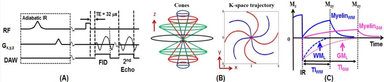
Two normal mice and two C57BL/6 mice six days post a standard controlled cortical impact (CCI) were sacrificed for this study. Brain imaging was performed on a Bruker 7T scanner. A mouse brain coil was used for signal reception. A 3D IR-UTE sequence was developed to image myelin with the following parameters: TR=1000 ms, inversion time (TI)=320 ms, TE=0.02 ms, FOV=2.2×2.2×2.2 cm3, matrix= 128×128×128 cm3, flip angle (FA)=20º. The acquired voxel size=172×172×172 µm3. To speed up data acquisition, a total number of spokes (Nsp) of 21 was acquired per IR preparation, leading to a total scan time of 100 min. Luxol Fast blue (LFB) staining was performed for assessment of demyelination. After the animal study, a 3D IR-UTE Cones sequence was developed on a GE MR750 3T scanner (Figure 1), and applied to five healthy volunteers and three mTBI patients. Informed consent was obtained from all subjects in accordance with guidelines of the institutional review board (IRB). The following sequence parameters were used: FOV=22×22 ×15.1 cm3, acquisition matrix=192×192×42, receiver bandwidth=250 kHz, TR/TI=1000/320 ms, TE=0.032ms/2.2ms, flip angle=20°, Nsp=21, and scan time=8.3 min. The 3D IR-UTE signal can be used to quantify myelin density by comparing myelin signal with that of a reference phantom. In this study, a rubber phantom was placed near the head for calibration9. Clinical T1- and T2-weighted FSE sequences were also used for comparison.Results and Discussion
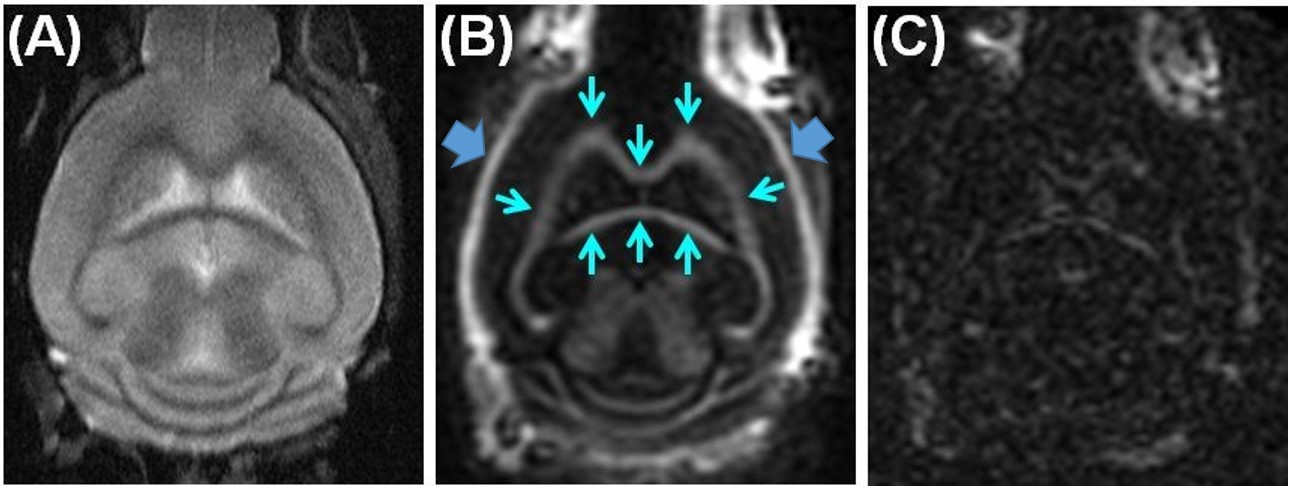
Figure 2 shows selected coronal slices from T2-FSE and 3D IR-UTE imaging of the normal C57BL/6 mouse Brain on a Bruker 7T scanner. Myelin was highlighted by the 3D IR-UTE sequence, but invisible with the T2-FSE sequence.
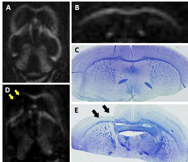
Figure 3 shows 3D IR-UTE imaging of a C57BL/6 mice three days post mTBI and of a normal control mouse. Obvious myelin loss was observed only in the mTBI mice, as confirmed on the LFB photomicrograph.
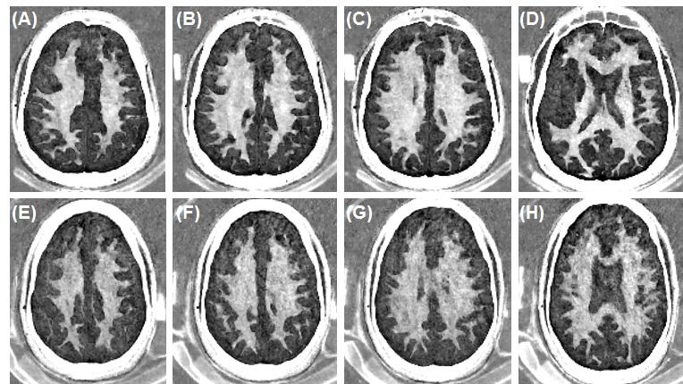
Figure 4 shows 3D IR-UTE imaging of myelin in white matter (WM) of the brain of a 27-year-old healthy male volunteer and a 29 year-old male mTBI patient, respectively. Volumetric imaging of myelin was achieved for both, demonstrating a myelin density of 7.8±0.7% for the healthy volunteer and reduced myelin density of 5.9±0.5% for the mTBI patient, suggesting partial myelin loss due to mTBI.
These preliminary results demonstrate the feasibility of volumetric myelin mapping using the 3D IR-UTE sequences at 7T and 3T. Further optimization of the technique may allow fast quantitative assessment of myelin relaxation times (T1 and T2*) and phase as well as proton density, providing a comprehensive assessment of myelin changes due to mTBI. This technique may significantly improve the diagnosis and treatment monitoring of mTBI patients.
Conclusion
Acknowledgements
The authors acknowledge grant support from GE Healthcare, NIH (R01NS092650), and the VA Clinical Science and Rehabilitation R&D Awards (I01CX001388 and I01RX002604)References
1. Mierzwa AJ, Marion CM, Sullivan GM, McDaniel DP, Armstrong RC. Components of myelin damage and repair in the progression of white matter pathology after mild traumatic brain injury. J Neuropathol Exp Neurol 2015; 74:218-232.
2. Johnston KM, Ptito A, Chankowsky J, Chen JK. New frontiers in diagnostic imaging in concussive head injury. Clin J Sport Med 2001; 11:166-175.
3. Flygt J, Djupsjo A, Lenne F, Marklund N. Myelin loss and oligodendrocyte pathology in white matter tracts following traumatic brain injury in the rat. EJN 2013; 38:2153-2165.
4. Mierzwa AJ, Marion CM, Sullivan GM, McDaniel DP, Armstrong RC. Components of myelin damage and repair in the progression of white matter pathology after mild traumatic brain injury. J Neuropathol Exp Neurol 2015; 74:218-232.
5. Ng HK, Mahaliyana RD, Poon WS. The pathological spectrum of diffuse axonal injury in blunt head trauma: assessment with axon and myelin stains. Clinical Neurology and Neurosurgery 1994; 96:24-31.
6. Liu MC, Akle V, Zheng W, Kitlen J, O’Steen B, Larner SF, Dave JR, Tortella FC, Hayes RL, Wang KKW. Extensive degradation of myelin basic protein isoforms by calpain following traumatic brain injury. J Neurochemistry 2006; 98:700-712.
7. Taib T, Leconte C, Steenwinchel JV, Cho AH, Palmier B, Torsello E, et al. Neuroinflammation, myelin and behavior: temporal patterns following mild traumatic brain injury in mice. PlosOne 2017; 12(9):e01847811.
8. Bigler ED, Maxwell WL. Neuropathology of mild traumatic brain injury: relationship to neuroimaging findings. Brain imaging and Behavior 2012; 6:108-136.
9. Du J, Ma G, Li S, Carl M, Szeverenyi N, VandenBerg S, Corey-Bloom J, Bydder GM. Ultrashort TE echo time (UTE) magnetic resonance imaging of the short T2 components in white matter of the brain using a clinical 3T scanner. NeuroImage 2013; 87C:32-41. PMID: 24188809.
10. Sheth V, Shao H, Chen J, VandenBerg S, Corey-Bloom J, Bydder GM, Du J. Magnetic resonance imaging of myelin using ultrashort echo time (UTE) pulse sequence: phantom, specimen, volunteers and multiple sclerosis patient studies. NeuroImage 2016; 136:37-44.
11. Sheth V, Fan S, He Q, Ma Y, Annesse J, Switzer R, Corey-Bloom J, Bydder GM, Du J. Inversion recovery UTE magnetic resonance imaging: a method for simultaneous direct detection of myelin and high signal demonstration of iron deposition in the brain–a feasibility study. Magn Reson Imaging 2017; 38:87-94.
12. Fan S, Ma Y, Zhu Y, Szeverenyi N, Bydder GM, Du J. Yet more evidence that myelin protons can be directly imaged with ultrashort echo time (UTE) sequences on a clinical 3T scanner: bi-component T2* analysis of native and deuterated ovine brain specimens. Magn Reson Med 2017; doi:10.1002/mrm.27052.
Figures