0111
Echo Planar Time-resolved Imaging (EPTI)1Athinoula A. Martinos Center for Biomedical Imaging, Massachusetts General Hospital, Charlestown, MA, United States, 2Harvard-MIT Health Sciences and Technology, MIT, Cambridge, MA, United States, 3Department of Electrical Engineering and Computer Science, MIT, Cambridge, MA, United States, 4Department of Radiology, Harvard Medical School, Boston, MA, United States
Synopsis
A new technique, termed Echo Planar Time-resolved Imaging (EPTI), was developed to address EPI’s geometric distortion and blurring, and to provide temporal signal evolution information across the EPI readout window. Using a small number of EPTI-shots, a time-series of multi-contrast images can be created free of distortion and blurring (up to 100 T2- and T2*-weighted images). This should make EPTI useful for numerous applications. Here, we demonstrated EPTI in brain to provide i) rapid simultaneous quantitative mapping of T2, T2*, proton density and tissue phase, as well as ii) multi-echo and quantitative T2* fMRI.
Introduction
Echo planar imaging (EPI)[1] is a popular fast MR acquisition technique, but suffers from a number of drawbacks including i) geometric distortion due to B0-inhomogeneity, ii) spatial blurring from T2/T2* decay, and iii) limited number of echoes in multi-echo imaging due to lengthy sequential readouts. These problems compromise EPI’s image quality in functional/diffusion/perfusion imaging, limit its application to anatomical imaging and preclude multi-echo EPI from common use for quantitative/multi-contrast imaging.
In this study, a new multi-shot EPI technique, termed Echo Planar Time-resolved Imaging (EPTI)[2], was developed to address these issues. This approach not only achieves distortion- and blurring-free imaging, it also provides up to 100 T2&T2*-weighted echo images across the EPI readout window, spaced at a time interval equal to EPI’s echo-spacing (~1ms). This should make it useful to numerous applications where high-SNR undistorted images or multiple-contrast images are desired. Here, we demonstrated EPTI for two in vivo brain applications: i) a combined gradient- and spin-echo EPTI for rapid quantitative mapping of T2, T2*, proton density, and susceptibility (1.1 mm in-plane resolution at 0.8 s/slice); and ii) a gradient-echo EPTI, for quantitative T2* whole-brain fMRI at 2×2×3 mm3 spatial and 3.3s temporal resolution.
Methods
To understand how EPTI works, a ky-t space of EPI signal is introduced in Fig.1. In conventional single-shot EPI (ss-EPI), the signal is acquired to fill a 45° diagonal line in the ky-t space, with T2/T2* decay and susceptibility-induced phase accumulating over time, leading to blurring and distortion in the final image. To correct for distortion, a pair of datasets with reversed phase-encoding can be acquired[3]. Such acquisition obtains two +/-45° diagonal lines in the ky-t space, with more information to estimate and correct for the susceptibility-induced distortion (Fig.1A). To obtain multiple-contrast images, multi-echo EPI methods[4,5] can be used as shown in Fig.1B, but suffer from limited number of echoes as well as image distortions and blurring.
If the ky-t space is fully sampled, distortion- and blurring-free images with different contrasts can be obtained at different echo times with a time interval of an echo-spacing. Such full ky-t coverage is however extremely encoding intensive, and EPTI aims to achieve this by using only a small number of EPTI-shots with a new highly-accelerated spatio-temporal CAIPI sampling strategy. As shown in Fig.1C, each EPTI shot covers a segment of the ky-t space using a zig-zag trajectory that contains multiple diagonal ky line-sections. The temporally adjacent ky line-sections are interleaved with complementary PE sampling. This trajectory ensures that neighboring ky-points within each EPTI shot are acquired only a few milliseconds apart, and contain small B0-inhomogeneity-induced phase and T2* decay that can be well estimated by our B0-inhomogeneity-informed parallel imaging reconstruction[2,6]. Compact kernels are used in the reconstruction to interpolate under-sampled ky-t space to fully-sampled ky-t by exploring signal correlation across time and coil dimensions. With this ky-t encoding view, EPTI readouts can be applied continuously within the sequence (Fig. 1C bottom) to fill any dead time, leading to high acquisition and SNR efficiency.
To further accelerate EPTI, simultaneous multi-slice (SMS)[7,8] was incorporated using an optimized ky-kz-t CAIPI trajectory. To provide robustness to shot-to-shot B0-variations, a navigation-free B0-variation estimation and correction method was also developed[2].
Results
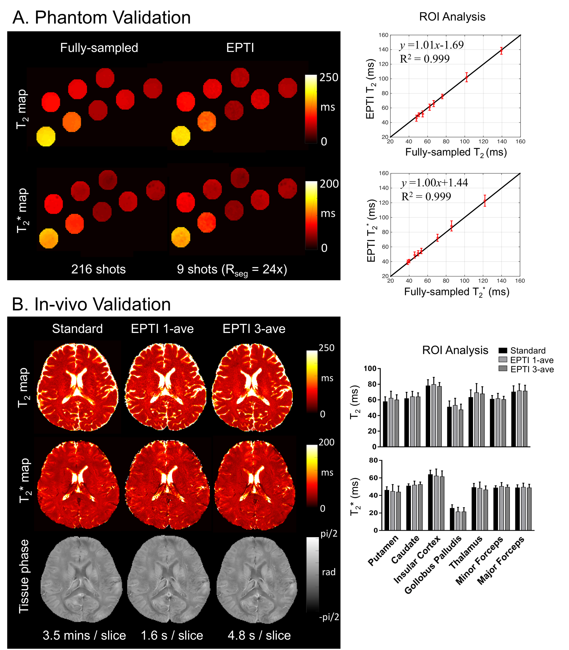
Fig.2 shows strong agreements in geometric brain boundaries of EPTI data and lengthy standard structural data, while ss-EPI results in severe distortions. Fig.3 shows high agreements in T2 and T2* maps obtained from lengthy gold standard acquisition and highly accelerated GE-SE EPTI acquisition (Rseg=24) in both phantom and in vivo. Fig.4 shows the results from 48× accelerated GE-SE EPTI where SMS has been incorporated (Rseg×MB = 24×2). Multi-contrast images and quantitative maps can be obtained in just ~0.8s per imaging slice at 1.1 mm in-plane resolution. Fig.5 shows preliminary results of using GE EPTI for a visual stimulation task fMRI experiment, where distortion-free whole-brain data at 2×2×3 mm3 resolution across 34 echoes can be obtained at a TReff of 3.3s.Discussion and Conclusion
The use of highly-accelerated spatio-temporal CAIPI trajectory and continuous EPTI readouts with no dead-time in acquisition have enabled more than 100 of multi-contrast images to be obtained in < 1s per imaging slice. Incorporation of SMS has also provided further boost in SNR-efficiency. Applications of the EPTI concept to 3D sequences and subspace-constrained reconstructions are underway to allow even higher SNR-efficiency and acceleration[9,10], while a navigation-free motion-robust EPTI acquisition is also being developed[11]. Highly-efficient continuous scan achieved through EPTI should provide a new approach to push the boundary of MRI encoding and speed, with many potential applications.Acknowledgements
This work was supported by NIH NIBIB (R01-EB020613, R01-EB019437, R01-MH116173, P41-EB015896 and U01-EB025162) and by the MGH/HST Athinoula A. Martinos Center for Biomedical Imaging; and was made possible by the resources provided by NIH Shared Instrumentation Grants S10-RR023401, S10-RR023043, and S10-RR019307.References
1. Mansfield P. Multi-planar image formation using NMR spin echoes. Journal of Physics C: Solid State Physics 1977;10(3):L55.
2. Wang F, Dong Z, Reese TG, Bilgic B, Katherine Manhard M, Chen J, Polimeni JR, Wald LL, Setsompop K. Echo planar time‐resolved imaging (EPTI). Magnetic resonance in medicine 2019.
3. Andersson JL, Skare S, Ashburner J. How to correct susceptibility distortions in spin-echo echo-planar images: application to diffusion tensor imaging. Neuroimage 2003;20(2):870-888.
4. Posse S, Wiese S, Gembris D, Mathiak K, Kessler C, Grosse-Ruyken M-L, Elghahwagi B, Richards T, Dager SR, Kiselev VG. Enhancement of BOLD-contrast sensitivity by single-shot multi-echo functional MR imaging. Magnetic resonance in medicine 1999;42(1):87-97.
5. Schmiedeskamp H, Straka M, Newbould RD, Zaharchuk G, Andre JB, Olivot JM, Moseley ME, Albers GW, Bammer R. Combined spin‐and gradient‐echo perfusion‐weighted imaging. Magnetic resonance in medicine 2012;68(1):30-40.
6. Dong Z, Wang F, Reese TG, Manhard MK, Bilgic B, Wald LL, Guo H, Setsompop K. Tilted‐CAIPI for highly accelerated distortion‐free EPI with point spread function (PSF) encoding. Magnetic resonance in medicine 2019;81(1):377-392.
7. Larkman DJ, Hajnal JV, Herlihy AH, Coutts GA, Young IR, Ehnholm G. Use of multicoil arrays for separation of signal from multiple slices simultaneously excited. Journal of Magnetic Resonance Imaging 2001;13(2):313-317.
8. Setsompop K, Gagoski BA, Polimeni JR, Witzel T, Wedeen VJ, Wald LL. Blipped‐controlled aliasing in parallel imaging for simultaneous multislice echo planar imaging with reduced g‐factor penalty. Magnetic Resonance in Medicine 2012;67(5):1210-1224.
9. Wang F, Dong Z, Reese TG, Wald LL, Setsompop K. 3D-EPTI for ultra-fast multi-contrast and quantitative imaging. 2019.
10. Dong Z, Wang F, Reese TG, Bilgic B, Setsompop K. Echo Planar Time-Resolved Imaging (EPTI) with Subspace Constraint and optimized k-t trajectory. 2019.
11. Fair MJ, Wang F, Dong Z, Bilgic B, Reese T, Setsompop K. Propeller Echo-Planar Time-resolved Imaging with Dynamic Encoding (PEPTIDE).
Figures