0110
SPARKLING: variable-density k-space filling curves for accelerated MRISynopsis
This work reports the use of new non-Cartesian k-space trajectories whose improved efficiency allows to significantly reduce MR scan time with minimum deterioration of image quality. Instead of using simple geometrical patterns, we introduce an approach inspired from stippling techniques, which automatically designs optimized sampling patterns along any desired density by taking full advantage of the hardware capabilities. We use our strategy to accelerate the acquisition time of T2*-weighted scans acquired at 7T on in vivo human brains. We compare our method to standard non-Cartesian trajectories (spiral, radial) and demonstrate its superiority regarding image quality and robustness to system imperfections.
Introduction
Reducing scan times in Magnetic Resonance Imaging (MRI) is essential to explore higher spatial resolution which could help to better diagnose certain pathologies such as Alzheimer's disease. Methods to accelerate MRI such as parallel imaging [1] or compressed sensing [2] commonly rely on simple sampling patterns such as straight lines, spirals or slight variations of these elementary shapes. Such strategies may not take full advantage of the degrees of freedom offered by the hardware. In addition, they cannot be easily adapted to fit arbitrary sampling distribution and may lack robustness at high acceleration factors (AF). Here, we report the use of a method called SPARKLING (Spreading Projection Algorithm for Rapid K-space sampLING), which may overcome these limitations by taking a completely different approach to the design of faster k-space sampling [3]. A key ingredient underlying our approach is to take advantage of a shorter dwell time to acquire more points in a given observation window. In addition, this method relies on optimization to automatically generate non-Cartesian k-space trajectories under the hardware constraints while fulfilling key criteria for optimal sampling: a controlled distribution (e.g. along a variable density) and a locally uniform coverage. To illustrate SPARKLING advantages over standard non-Cartesian sampling, we used the proposed sampling patterns to accelerate T2*-weighted 2D in vivo brain acquisitions and compared them with equally accelerated variable-density spiral and radial acquisitions. These data were prospectively acquired with a 7T scanner for high in plane resolution (390 μm x 390 μm) and various acceleration factors up to 20.Materials and Methods
Setup
Four healthy volunteers were scanned with a 7T system (Siemens Healthineers, Erlangen, Germany) and a 1Tx/32Rx head coil (Nova Medical, Wilmington, MA, USA). Maximum gradient and slew rate magnitudes on this scanner were respectively 40 mT/m and 333 T/m/s and the gradient raster time was 10 μs. All subjects signed a written informed consent form and was enrolled in the study under the approval of our institutional review board.
Sequence and k-space trajectoriesA modified 2D T2*-weighted GRE sequence was acquired for an in plane resolution of 390 μm with the following parameters: TR = 550 ms, TE = 30 ms and FA = 25° for 11 interleaved transversal slices of 3 mm. Acquisitions were performed using the SPARKLING trajectories for different numbers of shots (64, 51, 34 and 26). (Fig. 1) displays an accelerated SPARKLING trajectory segmented into 34 symmetric shots each acquiring 3072 samples during a readout time of 30.72 ms. The SPARKLING sampling was distributed along a radially decaying density. Limits in gradient and slew rate amplitudes were respectively set to 40 mT/m and 200 T/m/s. Moreover, we acquired a set of images with a variable-density in-out spiral [4] and a radial trajectory with the same numbers of shots, samples and acquisition time (TA) as SPARKLING patterns. The comparison involved 26-shot trajectories corresponding to TA = 14 s for 11 slices, which is 20 times faster than the fully-sampled acquisition of 512 Cartesian lines (TA = 4 min 42s).
Image reconstruction
Images were reconstructed using a self-calibrating nonlinear algorithm minimizing a sparsity promoting regularized CS-SENSE (Compressed Sensing SENsitivity Encoding) criterion in the wavelet domain similar to [5,6] which was adapted to non-Cartesian samples using the NFFT [7].
Results
(Fig. 2) shows the brain images of two different subjects for an axial orientation in (Fig. 2a‐c) and (Fig. 2d‐f) and of a third subject for a coronal orientation in (Fig. 2g). The axial reference scan in (Fig. 2a&d) lasted 4 min 42 s for 11 slices, while the acquisition times of the 8‐fold and 15‐fold accelerated SPARKLING scan were 35 s and 18 s, respectively. The high target image quality was fairly well maintained even for the 34‐shot SPARKLING trajectory. (Fig. 2g) displays a set of multiple coronal slices acquired with a SPARKLING trajectory composed of 51 shots, lasting 28 s (AF = 10). Consistent contrast and level of detail were obtained in all 11 slices.
The results from a fourth subject are presented in (Fig. 3) for the highest studied acceleration factor, AF = 20, corresponding to 26 shots. (Fig. 3b&e) shows the image reconstructed from the SPARKLING acquisition lasting 14 s for 11 slices. Compared to the reference in (Fig. 3a), which was obtained in an acquisition time of 4 min 42 s, the SPARKLING result was able to maintain the image quality fairly well despite a slight loss of resolution visible on the smallest vessels. However, the spiral acquisition at the same acceleration factor (Fig. 3c&g) has notably more artifacts, and the 20‐fold‐accelerated radial reconstruction (Fig. 3d&h) appears blurry and presents streaking artifacts.
Discussion and conclusion
We demonstrated the in vivo superiority of the proposed SPARKLING sampling over standard Cartesian, radial and spiral approaches for long-readout T2*-weighted imaging. Although accelerated by a factor of 20, SPARKLING acquisitions were able to preserve fine structural details and proved to be more robust to system imperfections. Most interestingly, our method can adapt to any desired sampling density, observation window, MR weighting and hardware specifications. Although we focused on 2D acquisitions as a proof-of-concept, our technique can be extended to 3D acquisitions [8] as well as to lower field MRI.
Acknowledgements
We would like to thank Nicolas Boulant and Vincent Gras for their insightful remarks. This research program was supported by DRF Impulsion grant in 2016 (COSMIC, P.I.: P.C.). C.L. was also supported by the CEA international PhD program.References
[1] : Pruessmann KP, Weiger M, Scheidegger MB, Boesiger P. (1999). SENSE: sensitivity encoding for fast MRI. Magnetic resonance in medicine. 42(5):952-62.
[2] : Lustig M, Donoho D, Pauly JM. (2007). Sparse MRI: The application of compressed sensing for rapid MR imaging. Magnetic resonance in medicine. 58(6):1182-95.
[3] : Boyer C, Chauffert N, Ciuciu P, Kahn J, Weiss P. (2016). On the generation of sampling schemes for Magnetic Resonance Imaging. SIAM Imaging Science, Vol. 9, N.4, 2039-2072.
[4] : Lee JH, Hargreaves BA, Hu BS, Nishimura DG. (2003). Fast 3D imaging using variable‐density spiral trajectories with applications to limb perfusion. Magnetic resonance in medicine. 50(6):1276-85.
[5] : Wu B, Millane RP, Watts R, Bones P. (2008). Applying compressed sensing in parallel MRI. Proceedings of the 16th Annual Meeting of ISMRM (Vol. 1480).
[6] : Liu B, Zou YM, Ying L. (2008). SparseSENSE: application of compressed sensing in parallel MRI. Information Technology and Applications in Biomedicine. ITAB 2008. International Conference (pp. 127-130).
[7] : Keiner, J., Kunis, S., and Potts, D. (2009). Using NFFT 3 - a software library for various nonequispaced fast Fourier transforms. ACM Trans. Math. Software, 36 : Article 19, 1-302.
[8]: Lazarus C, Weiss P, Chauffert N, Mauconduit F, Vignaud A, Ciuciu P. (2019). 3D SPARKLING for accelerated ex vivo T2*-weighted MRI with compressed sensing. 27th annual meeting of the International Society for Magnetic Resonance Imaging.
Figures
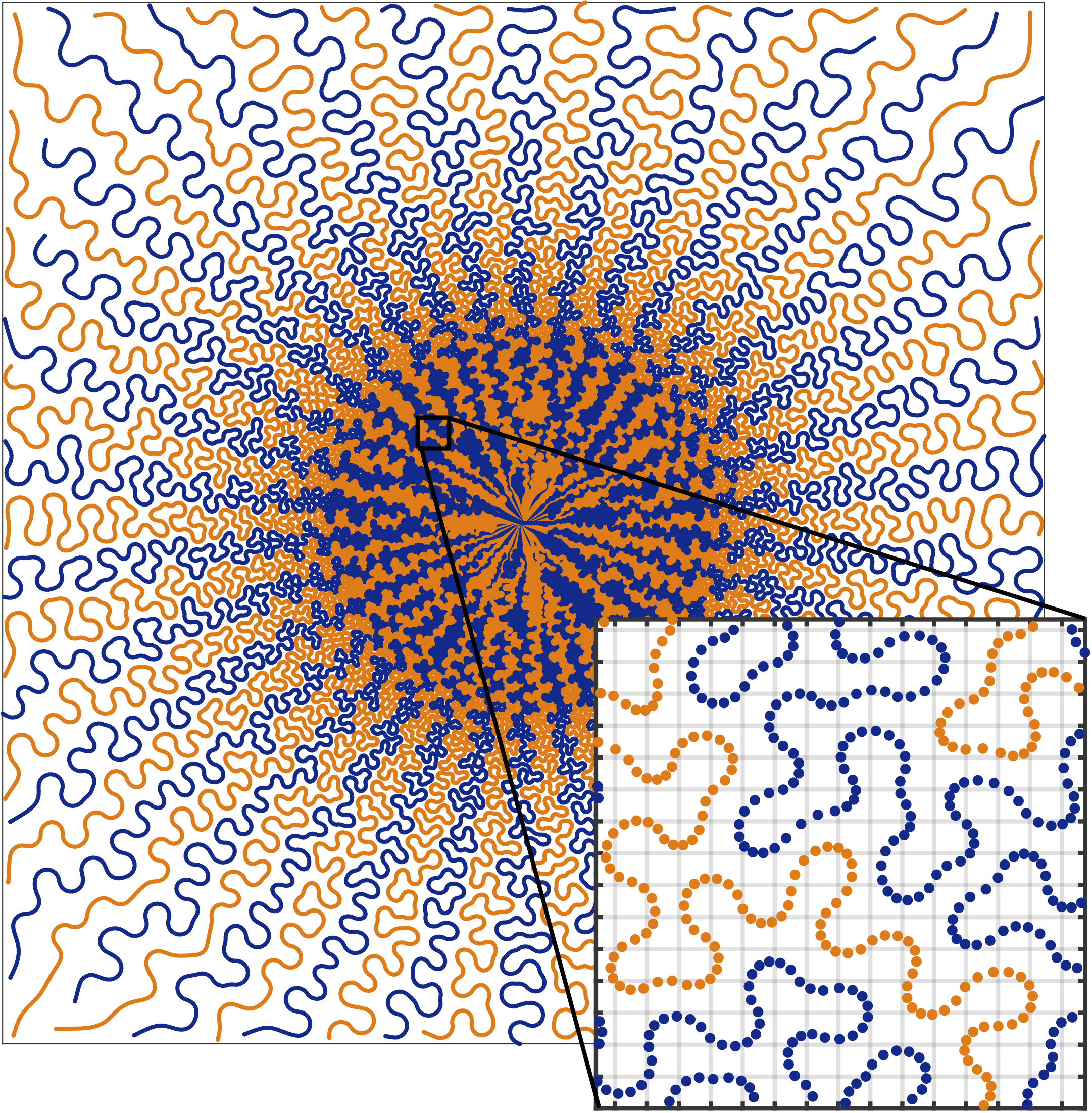
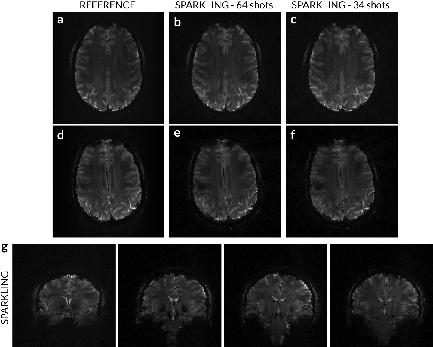
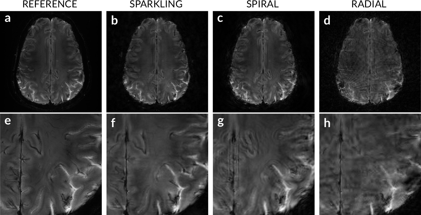
Figure 3: In vivo validation of radial‐initialized SPARKLING trajectories composed of 26 shots (AF = 20) and comparison with spiral and radial sampling. T2*‐weighted GRE acquisition on a 7 Tesla scanner at an image resolution of 390 μm x 390 μm x 3 mm. a, e, Fully sampled Cartesian reference with an acquisition time of 4 min 42 s for 11 slices and a magnified region of interest in the parieto‐occipital cortex. b, f, Image and magnified image reconstructed from a 20‐fold‐accelerated variable‐density SPARKLING acquisition lasting 14 s for 11 slices. c, g, Image and magnified image reconstructed from a 20‐fold accelerated variable‐density spiral acquisition lasting 14 s. d, h, Image and magnified image reconstructed from a 20‐fold accelerated radial acquisition lasting 14 s. Image reconstructions did not include any correction of system imperfections.