0109
Prediction of Peripheral Nerve Stimulation Thresholds of MRI Gradient Coils using Coupled Electromagnetic and Neurodynamic Simulations1Computer Assisted Clinical Medicine, Medical Faculty Mannheim, Heidelberg University, Heidelberg, Germany, 2Dept. of Radiology, A.A. Martinos Center for Biomedical Imaging, Massachusetts General Hospital, Charlestown, MA, United States, 3Harvard Medical School, Boston, MA, United States, 4Siemens Healthcare, Erlangen, Germany, 5Harvard-MIT Division of Health Sciences Technology, Cambridge, MA, United States
Synopsis
Peripheral Nerve Stimulation (PNS) has become the major limitation in many fast MRI sequences for state-of-the-art gradient systems. We present the first (to our knowledge) full PNS model for assessing magnetostimulation thresholds and a method to incorporate these thresholds as constraints in the coil-winding design phase. Our model consists of comprehensive female and male body models for EM simulations, co-registered atlases of peripheral nerves, and a neurodynamic model describing the nerve responses to induced electric fields. We validated our framework based on three commercial MR gradient systems and found close resemblance between simulated thresholds and experimentally obtained group PNS thresholds.
Purpose
Rapid switching of MR gradient coils induces electric fields in the human body strong enough to induce peripheral nerve stimulation (PNS)1,2. The occurrence of PNS often limits the usable performance of the gradient system, leading to longer scan-times or reduced spatial and temporal resolution3. Despite its impact, PNS metrics are only indirectly addressed during the coil design phase, e.g., by reducing the linear volume4 or by conducting stimulation experiments using healthy human subjects on coil prototypes. We developed the first framework to fully model PNS and predict stimulation thresholds and sites in the whole body, allowing predictions from hypothetical coil layouts without constructing expensive coil prototypes and facilitating development of other PNS mitigation strategies5,6. We show that the PNS estimates can be pre-computed for stream-function basis sets, allowing rapid determination of a PNS constraint during design optimization with the standard Boundary Element Method Stream Function (BEM-SF) approach7.Methods
PNS Simulation Framework: Our PNS modeling framework has three components: 1) detailed male and female surface body models for simulation of the induced EM-fields (Fig. 1B); 2) comprehensive atlases of peripheral nerves co-registered to the body models; each nerve labeled by the axon diameter; 3) a neurodynamic model of myelinated mammalian nerves (MRG model8,9) to predict the nerve’s response to the imposed E-fields (including generation of action potentials, APs). After simulation of the induced E-fields using a magneto quasi-static solver (Sim4Life, Zurich MedTech), we calculate the resulting electric potential changes along the nerves by projecting the E-field onto the nerves and integration. These potentials change both spatially along the nerves and temporally (the applied current waveform). The PNS threshold for a given coil waveform is determined by iteratively increasing the waveform amplitude until an AP is generated in any nerve. Threshold curves are plotted as the minimum stimulating gradient amplitude as a function of the gradient rise-time (“pulse duration”). Gradient Coils: We simulated PNS thresholds for two Siemens body gradients and one head gradient (Fig. 1A) and compared the simulated thresholds with the known experimental thresholds (averaged over 65 healthy adult subjects). Both simulations and experiments used sinusoidal and trapezoidal ramp times between 100us and 1000us (with constant 500us flat top duration) played in both single axis and combined axes (“X+Y”) gradient operation modes.Results
Figure 2 summarizes our PNS simulation workflow, starting with the E-fields in the male arm (outlines indicate tissue boundaries), including an exemplary nerve fiber path (dots). Projection and integration of the E-field along the nerve yields the electric potential (Fig. 2B). Figure 2C shows the neural activation function, NAF10 (defined as the second spatial derivative of the potential), an indicator of where the nerve is being stimulated. Excitation of this nerve fiber with a sinusoidal waveform and evaluation of the MRG neurodynamic model yields the membrane dynamics shown in Fig. 2D (plotted as the nerve’s transmembrane potential difference as a function of location along the nerve and time). Four APs are clearly visible in the membrane dynamics.
Figure 3 shows maps of the PNS oracle11 for BG1 and HG1 in the male body. This metric is similar to the NAF, but is adjusted to better correlate with the PNS thresholds. BG1 primarily stimulates nerves in the shoulders via the Y-axis, whereas HG1 stimulates the facial nerves via the X-axis (which correlates well with experimental observations). Figure 4 shows experimental thresholds (blue, incl. SD over all subjects) and simulated thresholds (female, male, and average between genders). There is good agreement between the average experimental and simulated thresholds: for all axes, axes combinations, and coil waveforms the NRMSE was ≤ 5% for BG1 and BG2 (not shown here, see [6]), and ≤ 10% for HG1.
Conclusion
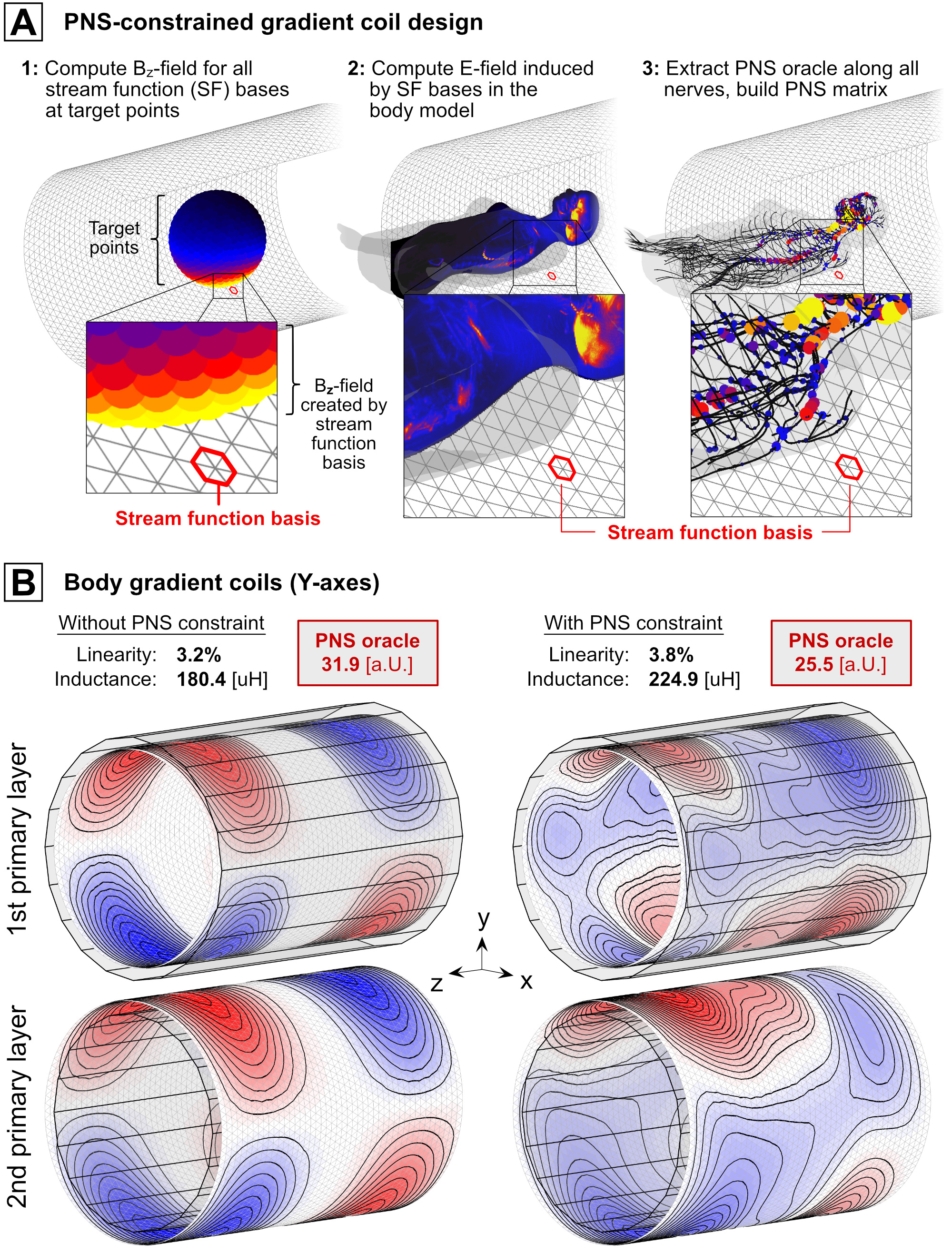
We demonstrate that PNS thresholds of MR gradient coils can be accurately predicted using coupled electromagnetic and neurodynamic simulations. Our PNS modeling tool can be used to study the mechanisms behind PNS and to identify degrees-of-freedom in MR gradient encoding that can be translated to PNS mitigation. We have recently extended the standard BEM-SF coil design approach12,13 to include the PNS oracle as a constraint. The PNS oracle can quickly compute PNS thresholds for a trial winding pattern as a linear combination from the stream function bases (see Fig. 5). In addition to introducing PNS as an explicit constraint in the optimization process (alongside traditional constraints such as gradient efficiency, linearity, inductance, and torque), this approach allows evaluating the tradeoffs between PNS and these constraints.Acknowledgements
The authors would like to acknowledge the help of past and present members of the gradient coil group at Siemens Healthineers, including Eva Eberlein, Peter Dietz, Ralph Kimmlingen, and Franz Hebrank. Research reported in this publication was supported by National Institute of Biomedical Imaging and Bioengineering, and the National Institute for Mental Health of the National Institutes of Health under award numbers R24MH106053, R00EB019482, U01EB025121, and U01EB025162. The content is solely the responsibility of the authors and does not necessarily represent the official views of the National Institutes of Health.References
[1] Mansfield et al., “Limits to neural stimulation in echo-planar imaging”. Magnetic Resonance in Medicine; 29:746–758, 1993
[2] Irnich et al., “Magnetostimulation in MRI”. Magnetic Resonance in Medicine; 33:619–623, 1995
[3] Setsompop et al., “Pushing the limits of in vivo diffusion MRI for the human connectome project”. NeuroImage; 80, 2013
[4] Zhang et al., “Peripheral nerve stimulation properties of head and body gradient coils of various sizes. Magnetic Resonance in Medicine”, 50, 2003
[5] Davids et al., “Predicting magnetostimulation thresholds in the peripheral nervous system using realistic body models”, Sci. Rep. 7:5316, 2017
[6] Davids et al., “Prediction of peripheral nerve stimulation thresholds of MRI gradient coils using coupled electromagnetic and neurodynamic simulations”. Magnetic Resonance in Medicine; 81(1), 2018
[7] Davids et al., “Peripheral Nerve Stimulation (PNS) constrained gradient coil design within a Boundary Element Method Stream Function (BEM-SF) optimization”. ISMRM 2019
[8] McIntyre et al., “Modeling the excitability of mammalian nerve fibers: Influence of afterpotentials on the recovery cycle”. J Neurophysiol. 87(2), 2002
[9] Richardson et al., “Modelling the effects of electric fields on nerve fibres: Influence of the myelin sheath”. IEEE Trans. Bio. Eng. 38(4), 2000
[10] Basser et al., “The activating function for magnetic stimulation derived from a three-dimensional volume conductor model”. Medical and Biological Engineering and Computing. 39(11), 1992
[11] Davids et al., “The PNS oracle: a modified activation function metric for rapid assessment of Peripheral Nerve Stimulation (PNS)”. ISMRM 2019
[12] Peeren et al., “Stream function approach for determining optimal surface currents”. Journal of Computational Physics, 191(1), 2003
[13] Lemdiasov et al., “A stream function method for gradient coil design”, Concepts Magn. Reson., 26(1), 2005
Figures