0100
Radiomics Analysis of tumor and peri-tumor tissue on T2-Weighted Imaging Improves Diagnostic Performance of Lymph Node Metastasis in Patients with Cervical Cancer1Department of Radiology, Henan Provincial People's Hospital, Zhengzhou, China, 2CAS Key Laboratory of Molecular Imaging, Institute of Automation, Beijing, China, 3School of Information and Electronics, Beijing Institute of Technology, Beijing, China, 4Cooperative Innovation Center of Internet Healthcare & School of Software and Applied Technology, Zhengzhou University, Zhengzhou, China
Synopsis
The tumor margin and peritumoral tissue play an important role in the process of LN metastasis. The aim of this study was to utilize radiomics analysis of tumor and peri-tumor tissue on T2 weighted image (T2WI) to improve LNM prediction ablility in cervical cancer patients. We found that peritumoral tissue of cervical cancer on T2WI showed favorable value in predicting LNM. The decision tree we proposed which incorporates the radiomics features of intratumoral and peritumoral tissue on T2WI and c-LN status can be potentially used for personalized preoperative evaluation of LNM and optimal treatment regimen selection in cervical cancer patients.
Background and Purpose
Prediction of lymph node metastasis (LNM) in locally advanced cervical cancer patients is of paramount importance for treatment regimen selection. Lymphatics in the tumor margin facilitate metastasis to regional lymph node (LN).1,2 The peritumoral tissue plays an important role in the process of LN metastasis. The aim of this study is to utilize radiomics analysis of tumor and peri-tumor tissue on T2 weighted image (T2WI) to improve diagnostic performance of LNM in cervical cancer patients.Materials and methods
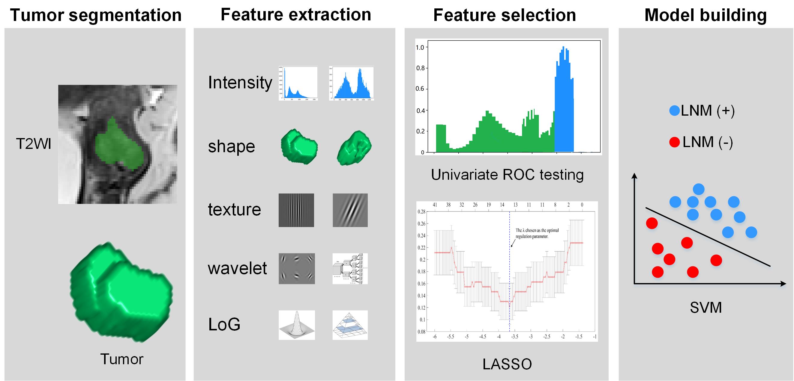
A total of 189 consecutive patients with cervical cancer who were treated between March 2012 and December 2017 were divided into a training cohort (n=126) and a validation cohort (n=63). All MRI scans were reviewed by two radiologists with 9 and 8 years of experience in pelvic disease interpretation. Based on commonly used criteria in daily clinical practice, patients with the short diameter of largest LN larger than 10mm were regarded as positive clinical LN (c-LN) status.3 For each patient, we extracted radiomics features from intratumoral and peritumoral tissues on sagittal T2WI. Afterwards, the radiomics features associated with LNM status were selected by univariate ROC testing and logistic regression with the least absolute shrinkage and selection operator (LASSO) penalty in the training cohort. Based on the selected features, a support vector machine (SVM) model was established to predict LNM status. The radiomics workflow is presented in Figure 1. To further improve the diagnostic performance, a decision tree which combines the radiomics model with clinical factors was built.Results
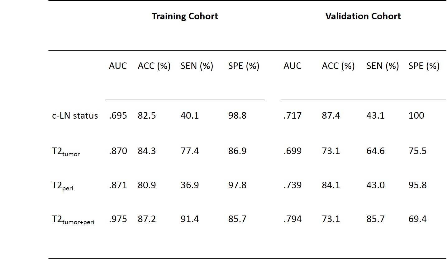
As shown in Figure 2, radiomics model of the intratumoral and peritumoral tissue on T2WI (T2tumor+peri) showed best sensitivity in detecting LNM, with 91.4% and 85.7% in the training and validation cohort respectively, and the c-LN status showed best specificity, with 98.8% and 100% in the training and validation cohort respectively, following by radiomics model of peritumoral tissue. Thus we proposed a decision tree for personalized evaluation of LNM. First, Radiomics model of T2tumor+peri was employed to evaluate if the patients have LNM, if LNM was considered high risk by the model, then we thought LNM was positive for this patient. If LNM was considered as low risk, c-LN status was employed to estimate if LNM did not exist. Patient with c-LN status negative was considered LNM negative and patient with c-LN status positive was considered LNM positive. The decision tree that combines radiomics model of T2tumor+peri and c-LN status achieved best diagnostic performance, the AUC, sensitivity, specificity were 0.895, 94.3% and 84.6%; 0.847, 100% and 69.3% in the training and validation cohorts respectively.Conclusions
The decision tree we built, which incorporates radiomics model of T2tumor+peri and c-LN status provides personalized evaluation of lymph node metastasis with high diagnostic performance and can be potentially applied in the preoperative prediction of LNM in locally advanced cervical cancer patients.Acknowledgements
This research was supported by the NNSFC (81720108021, 81772009,81601466,81641168, 31470047), National Key R&D Program of China (YS2017YFGH000397), Scientific and Technological Research Project of Henan Province (182102310162) and the Key Project of Henan Medical Science and Technology Project (201501011).References
1.Jain RK, Tong RT, Munn LL. Effect of vascular normalization by antiangiogenic therapy on interstitial hypertension, peritumor edema, and lymphatic metastasis: Insights from a mathematical model. Cancer Res. 2007;67(6):2729-2735.
2.Isaka N, Padera TP, Hagendoorn J, et al. Peritumor lymphatics induced by vascular endothelial growth factor-c exhibit abnormal function. Cancer Res. 2004;64(13):4400-4404.
3.Choi HJ, Roh JW, Seo SS, et al. Comparison of the accuracy of magnetic resonance imaging and positron emission tomography/computed tomography in the presurgical detection of lymph node metastases in patients with uterine cervical carcinoma: A prospective study. Cancer. 2006;106(4):914-922.
Figures