0099
Three-Dimensional Amide Proton Transfer MR Imaging for Cervical Cancer: Initial ExperienceYong-Lan He1, Cheng-Yu Lin1, Ya-Fei Qi1, Xiaoqi Wang2, Hai-Long Zhou1, Hua-Dan Xue1, and Zheng-Yu Jin1
1Peking Union Medical College Hospital, Beijing, China, 2Philips Healthcare China, Beijing, China
Synopsis
The present study demonstrates the feasibility of 3D APT MR imaging for uterine cervix on the largest sample size to our knowledge. As the clinical robustness of APT imaging in the pelvis is what researchers concerned, our study investigated and revealed the excellent agreement in both image quality assessment and APT values measurement, even on small cervical lesions with maximum diameter less than 2cm. Our studies demonstrated that APT imaging could differentiate cervical cancer from normal cervix. Cervical cancer showed significant higher APT values than that of normal cervix.
INTRODUCTION
Cervical cancer ranks as the fourth most frequently diagnosed cancer and the fourth leading cause of cancer death in women.1 According to NCCN Guidelines 2019, pelvic MR imaging is recommended to assess local disease extent in both non-fertility sparing and fertility sparing patients.2 Amide Proton Transfer (APT) MR imaging is a new contrast-agent-free MRI technique that addresses the need for a more confident diagnosis in oncology.3 The clinical utility of APT imaging for estimating the tumor of glioma, breast, lung cancers, prostate cancers, rectal cancer has been reported.4-8 We hypothesized that APT imaging might also be useful to estimate the lesion of uterine cervical cancer. The purpose of our study was to investigate the feasibility and clinical robustness of APT imaging for cervical cancer.METHODS
Institutional review board approval and informed consent were obtained. Between September 2017 and September 2018, 75 consecutive patients aged 24-74 years (mean age 45.6 years) pathologically confirmed of cervical lesions by colposcopy, and 49 normal volunteers, were scheduled for pelvic APT MR imaging on a 3T MR scanner (Ingenia 3.0T CX; Philips Healthcare, Best, the Netherlands) equipped with dual radiofrequency (RF) transmission subsystem. Two radiologists blindly evaluated APTw images quality by a 5 point Likert-scale on 64 patients with pathologically confirmed cervical cancer. APT values, calculated based on asymmetry of acquired Z-spectrum with respect to water frequency, using 3D TSE volume acquisition with B0 correction, were independently measured by two radiologists on 52 cervical cancer lesions and 49 normal cervical stroma with mean ROI area of 637.7 mm2 and 557.5 mm2 respectively. Inter-observer agreement was evaluated by Cohen’s Kappa analysis. Intra and inter-observer inter-class correlation coefficient (ICC) was computed. Student's t-test was performed to compare the differences of APT values between cervical cancer and normal cervix. ROC analysis was performed to computationally determine the feasible threshold value, sensitivity and specificity.RESULTS
Most cases revealed good APTw images quality ranked as score 4 with excellent agreement (к=0.823) (Fig. 1). Motion artifact was not significant in region of cervix. APT values of cervical cancer and normal cervical stroma were 2.745±0.065 and 1.853±0.059 respectively with statistical significant difference (p<0.0001, 95%CI 0.088-1.068) (Fig. 2).. Intra-observer ICC was 0.963 (95%CI 0.934-0.980) and 0.960 (95%CI 0.928-0.978) for two readers. Inter-observer ICC was 0.993 (95%CI 0.988-0.996). The AUC for differentiating cervical cancer from normal cervical stroma was 0.927. The feasible threshold value was determined as 2.221 with sensitivity of 84.62% and specificity of 83.66% (Fig. 3). For small lesions with the maximum diameter less than 2cm (n=15), the intra-observer ICC was 0.980 (95%CI 0. 941-0.994) and 0.985 (95%CI 0.954-0.995) for two readers. Inter-observer ICC was 0.990 (95%CI 0.971-0.997). For large lesions with the maximum diameter more than 2cm (n=37), the intra-observer ICC was 0.958 (95%CI 0. 915-0.980) and 0.952 (95%CI 0.902-0.976) for two readers. Inter-observer ICC was 0.994 (95%CI 0.989-0.997). Mean APT values of cervical cancer with the maximum diameter less than 2cm (2.698±0.116) showed no statistically significant difference compared with lesions more than 2cm (2.765±0.079) (p=0.648).DISCUSSION
CONCLUSION
APT MR imaging is feasible in cervical cancer detection. Cervical cancer showed significant higher APT values than that of normal cervix.Acknowledgements
This work was supported by grant from Beijing Natural Science Foundation (grant No. 7184234). The authors thank Zhang Pei and Meng Hua-Wei for their great help in this study.References
1. Bray F, Ferlay J, Soerjomataram I, Siegel RL, Torre LA, Jemal A (2018) Global cancer statistics 2018: GLOBOCAN estimates of incidence and mortality worldwide for 36 cancers in 185 countries. CA Cancer J Clin. 10.3322/caac.21492 2. Network NCC (2018) Clinical Practice Guidelines in Oncology. Cervical Cancer, Version I.2019. National Comprehensive Cancer Network, Plymouth Meeting. Available via https://www.nccn.org/professionals/physician_gls/pdf/cervical.pdf. Accessed 27 Sept 2018 3. Kamimura K, Nakajo M, Yoneyama T et al (2018) Amide proton transfer imaging of tumors: theory, clinical applications, pitfalls, and future directions. Jpn J Radiol. 10.1007/s11604-018-0787-3 4. Su C, Zhao L, Li S et al (2018) Amid proton transfer (APT) and magnetization transfer (MT) MRI contrasts provide complimentary assessment of brain tumors similarly to proton magnetic resonance spectroscopy imaging (MRSI). Eur Radiol. 10.1007/s00330-018-5615-8 5. Krikken E, Khlebnikov V, Zaiss M et al (2018) Amide chemical exchange saturation transfer at 7 T: a possible biomarker for detecting early response to neoadjuvant chemotherapy in breast cancer patients. Breast Cancer Res 20:51 6. Ohno Y, Kishida Y, Seki S et al (2018) Amide proton transfer-weighted imaging to differentiate malignant from benign pulmonary lesions: Comparison with diffusion-weighted imaging and FDG-PET/CT. J Magn Reson Imaging 47:1013-1021 7. Jia G, Abaza R, Williams JD et al (2011) Amide proton transfer MR imaging of prostate cancer: a preliminary study. J Magn Reson Imaging 33:647-654 8. Nishie A, Takayama Y, Asayama Y et al (2018) Amide proton transfer imaging can predict tumor grade in rectal cancer. Magn Reson Imaging 51:96-103 9. Law BKH, King AD, Ai QY et al (2018) Head and Neck Tumors: Amide Proton Transfer MRI. Radiology 288:782-790Figures
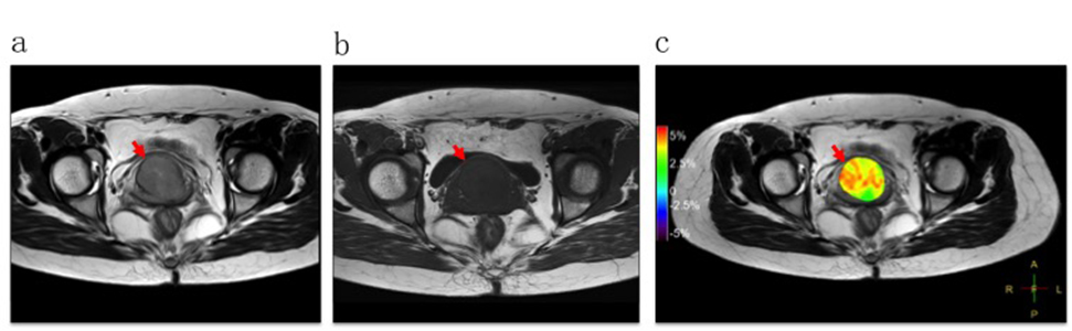
Fig. 1 A 43-year-old
woman with cervical squamous cell carcinoma. T2WI (a), T1WI (b) and APTw-T2WI
fusion image (c) showed the solid tumor on uterine cervix with maximum diameter
of 30.3m.
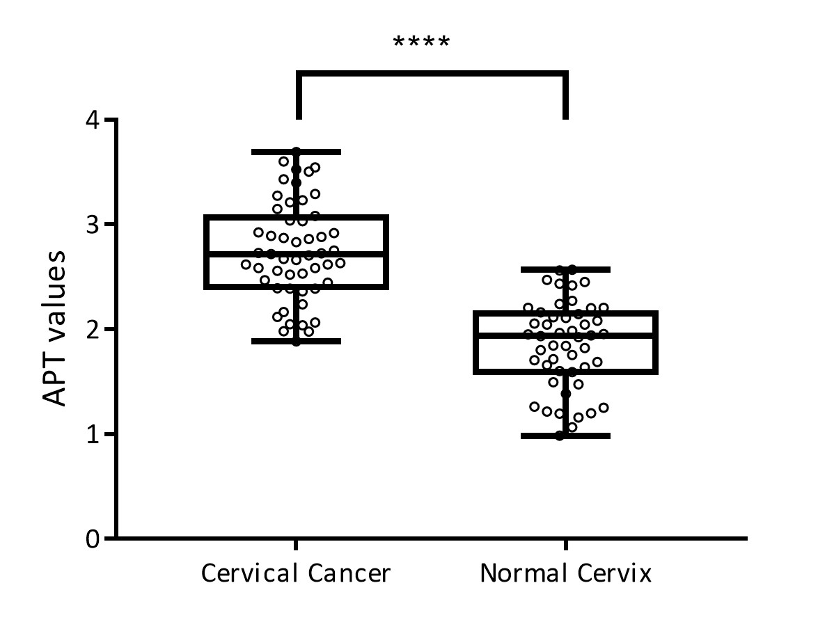
Fig. 2 APT values of
cervical cancer and normal cervical stroma were 2.745±0.065 and 1.853±0.059
respectively with statistically significant difference (p<0.0001).
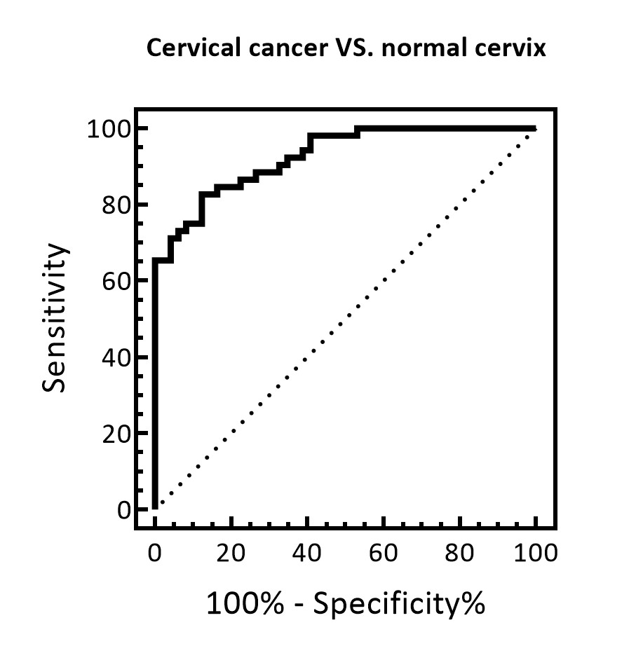
Fig. 3 ROC analysis for
differentiating cervical cancer from normal cervical stroma. Area under the
curve (AUC) was 0.927. The feasible threshold values was determined as 2.221
with sensitivity of 84.62% and specificity of 83.66%.