0097
Value of R2* and T2* in differential diagnosis of uterine sarcoma and degenerated hysteromyomaMiao Niu1, Ailian Liu1, and Lizhi Xie2
1The First Affiliated Hospital of Dalian Medical University, DaLian, China, 2GE Healthcare, MR Research, Beijing, China
Synopsis
To investigate the clinical value of enhanced T2 star weighted angiography (ESWAN) quantitative parameters in differential diagnosis of uterine sarcoma and degenerated hysteromyoma.
Purpose
To investigate the clinical value of enhanced T2 star weighted angiography (ESWAN) quantitative parameters in differential diagnosis of uterine sarcoma and degenerated hysteromyoma.Method and Materials
Result
The parameters measured by the two observers agreed with each other well(ICC>0.75). The R2* value of US was statistically lower than that of DH6.45(2.71,12.64)vs 15.20(13.08,17.03), p=0.000. The T2* value of US was statistically higher than DH 179.71(87.03,221.65)vs 6.37(59.51,81.60), P=0.000. The area under the ROC curve(AUC) of R2* and T2* was 0.928,0.862 respectively. The threshold of R2* and T2* for differentiating US from DH was ≥12.70、≤93.59 respectively and the sensitivity, specificity were 89.7%,84.5%、93.1%,76.9% respectively(Table2).Conclusion and Discussion
The R2* and T2* from ESWAN can effectively identify US and DH which can be a good choose to guide clinical treatment and assess prognosis. Uterine sarcoma (US) and degenerated hysteromyoma (DH) are two kinds of tumors of the female reproductive system, but prognosis is completely different. During to the similarity in images, they often misdiagnosed. So it is very important to distinguish them.Acknowledgements
No acknowledgement found.References
[1] DeMulder D, Ascher SM. Uterine Leiomyosarcoma: Can MRI Differentiate
Leiomyosarcoma From Benign Leiomyoma Before Treatment? AJR Am J Roentgenol. 2018
Oct 24:1-11.
[2] Takeuchi M, Matsuzaki K, Harada M. Clinical utility of susceptibility-weighted
MR sequence for the evaluation of uterine sarcomas. Clin Imaging. 2018 Oct
13;53:143-150.
Figures
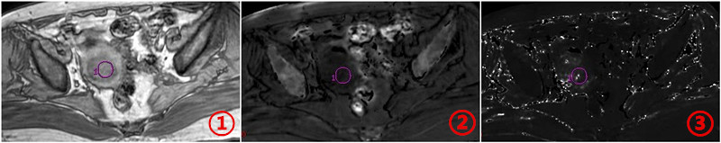
Figure.1(①-③)US ①ESWAN image ②R2* reconstruction
image (ROI placed in the center of the lesion, to avoid cystic degeneration,
necrosis, bleeding, get R2* value) ③T2* reconstruction image (ROI placed in the center of the
lesion, to avoid cystic degeneration, necrosis, bleeding, get T2* value).
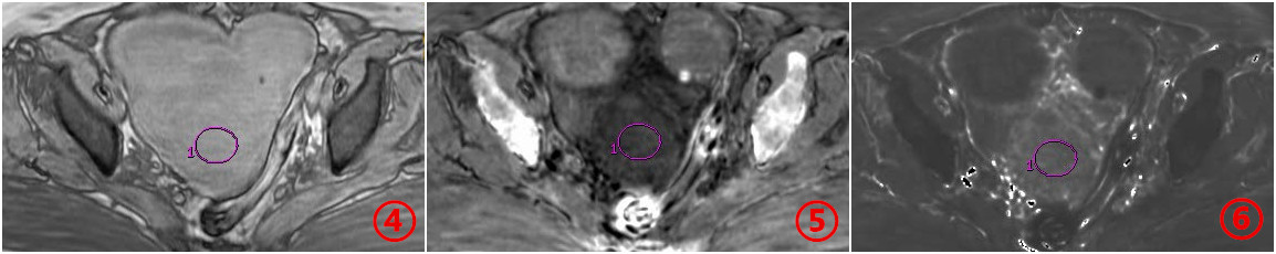
Figure.2(④-⑥)DH ④ESWAN image ⑤R2* reconstruction
image (ROI placed in the center of the lesion, to avoid cystic degeneration,
necrosis, bleeding, get R2* value) ③T2* reconstruction image (ROI placed in the center of the
lesion, to avoid cystic degeneration, necrosis, bleeding, get T2* value)
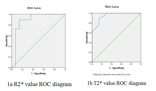
Figure.3 R2 * and T2 *
values to diagnose the ROC curves for US and DH.
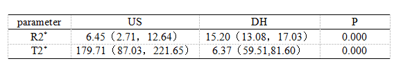
Table.1 Comparison of
the R2 * and T2 * values between US and DH.
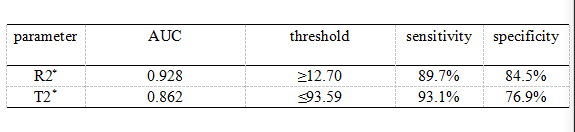
Table.2 The AUC,
threshold, sensitivity, and specificity of the R2 * and T2 * values of US and
DH.