0085
Changes In White Matter Microstructure In Relation To Working Memory After Mild Traumatic Brain Injury: Multi-Shell Diffusion MRI Study1Center for Advanced Imaging Innovation and Research (CAI2R), Department of Radiology, NYU School of Medicine, New York, NY, United States, 2Bernard and Irene Schwartz Center for Biomedical Imaging, NYU School of Medicine, New York, NY, United States, 3Department of Rehabilitation Medicine, NYU School of Medicine, New York, NY, United States
Synopsis
Working memory is a critical cognitive functions affected after mild traumatic brain injury (MTBI). We investigate associations between white matter (WM) microstructure and working memory, using multi-shell diffusion MRI and WAIS-IV subtests. The significant positive correlations observed in normal controls (NC) between tissue microstructure markers (fractional anisotropy (FA) and axonal water fraction (AWF)) with letter-number sequencing (LNS) were not present in MTBI. For MTBI, a significant positive correlation was observed between axial kurtosis (AK) and digit span backward (DSB), not seen in NC. These results show clear differences in the relationship between WM microstructure and working memory performance after injury.
INTRODUCTION
Mild traumatic brain injury (MTBI) is a significant public health problem,1 and at least 15% of patients report persistent cognitive complaints. The most common complaints and deficits in MTBI patients fall in the domain of working memory.2-3 Working memory is a system at the core of many cognitive functions and is responsible for holding, processing and manipulating information.4 Previous work shows that MTBI results in areas of white matter (WM) injury. Here we investigate how WM changes as assessed by multi-shell diffusion MRI relates to deficits in working memory post injury. Specifically we study the relationship between DTI, DKI and WM tract integrity (WMTI)5 metrics and Wechsler Adult Intelligence Scale-Fourth Edition (WAIS-IV)6 subtests tapping auditory-verbal working memory.METHODS
We studied 19 MTBI patients (age, 30±7, range 22-45yrs; 8 male) within a month of injury and 20 normal controls (NC) (mean age, 33±10, range 19-65yrs; 9 male). Non-native English speakers and non-right-handed individuals were excluded. Diffusion imaging was performed on a 3T MR scanner (Skyra, Siemens) with 5 b-values (0.25,1,1.5,2,2,2.5ms/µm2) along with 6,20,20,30,60 diffusion encoding directions using multiband (factor of 2) echo-planar imaging for accelerated acquisitions. Three images with b=0 were acquired. For geometric distortion correction, an additional image with b=0 was acquired with reversed phase encoding direction. Other parameters were: FOV=220×220mm2, matrix=88×88, resolution=2.5×2.5×2.5mm3, slices=56, TR/TE=4.9s/95ms, BW/pixel=2104Hz, a GRAPPA factor=2. We calculated maps of DTI metrics (fractional anisotropy [FA], mean, axial, radial diffusion coefficients [MD, AD, RD]) and DKI metrics (mean, axial, radial kurtosis [MK, AK, RK]) as well as WMTI metrics (axonal water fraction [AWF], intra-axonal diffusivity [Daxon], extra-axonal axial and radial diffusivities [De,a and De,r]). Auditory-verbal working memory was assessed using two WAIS-IV subtests: 1) Digit Span which includes Forward [DSF], Backward [DSB] and Sequencing [DSS]; and 2) Letter-Number Sequencing [LNS]. We performed tract-based spatial statistics (TBSS)7 and 27 WM regions-of-interest (ROIs) analyses with age/gender as covariates to reveal possible correlations between working memory subtest scores and diffusion metrics separately in NC and MTBI groups. For TBSS, statistical threshold level of p<0.05 was applied after family-wise error (FWE) correction for multiple comparisons. For ROI analysis, Pearson’s correlation test was performed in each ROI and Bonferroni correction was applied to adjust for multiple comparisons (p<0.0045). Correlation coefficients (R) were also calculated.RESULTS
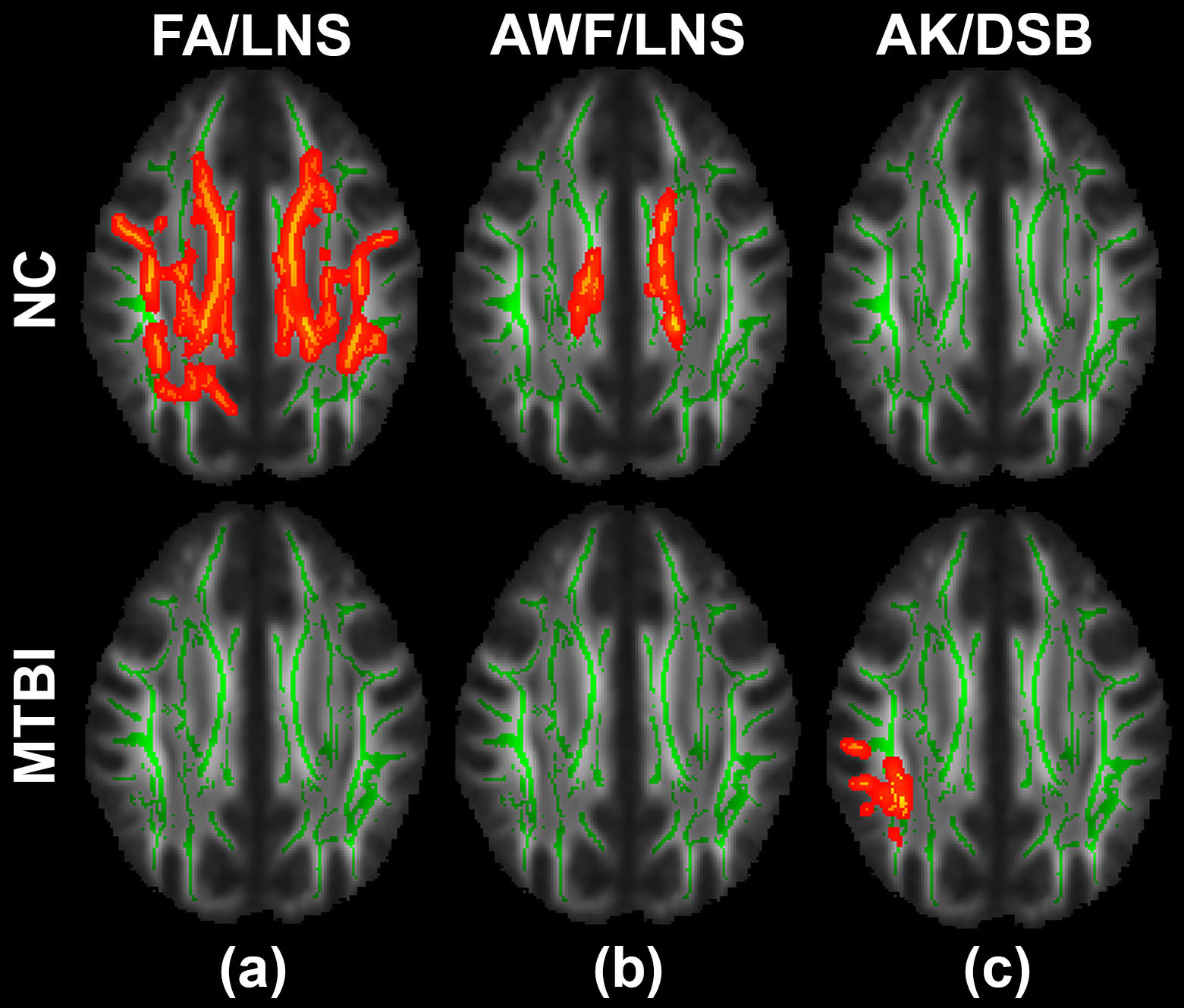
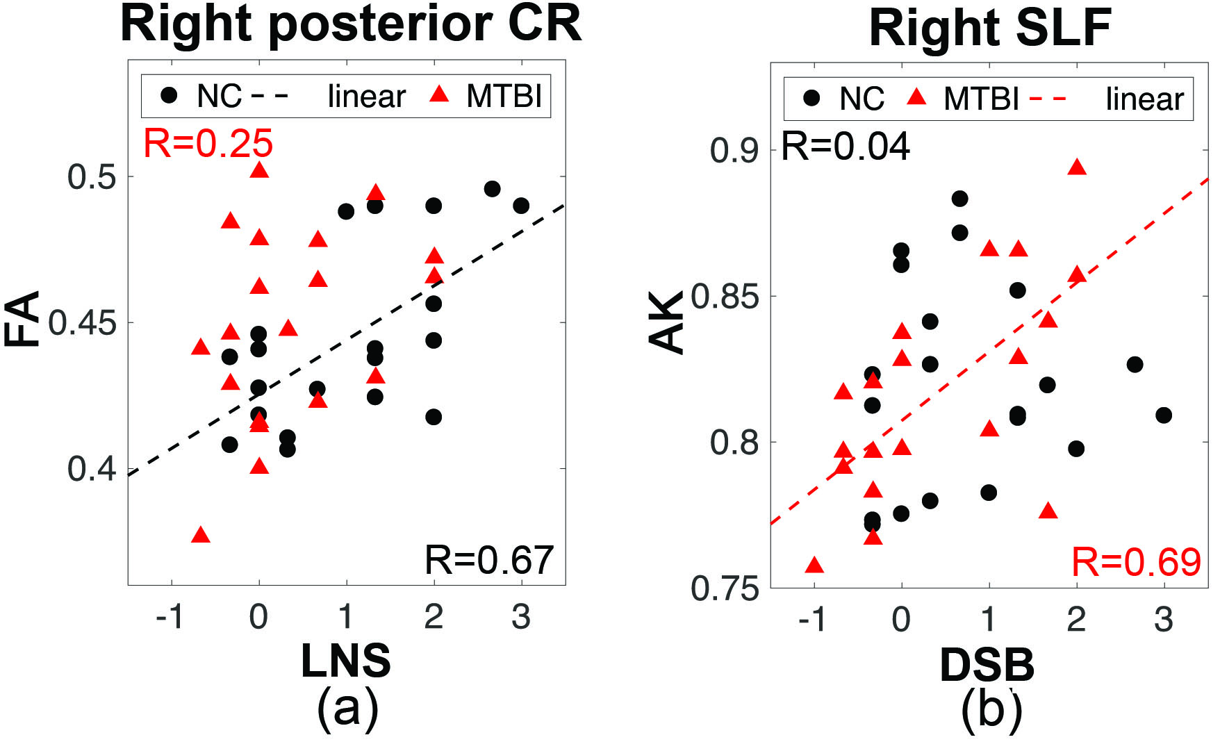
TBSS revealed multiple areas with statistically significant positive correlations between FA/AWF and LNS in NC, involving parietal WM, superior/posterior corona radiata (CR) and body/splenium of corpus callosum (CC), that were not present in the MTBI group (Fig.1(a-b)). In the MTBI group, a significant positive correlation was found between AK and DSB in the right superior longitudinal fasciculus (SLF) that was absent in NC (Fig.1(c)). From ROI analysis, significant positive correlations were found between FA and LNS in right posterior CR in NC (R=0.67,p=0.002), not present in the MTBI group (Fig.2(a)); and between AK and DSB in right SLF in the MTBI group (R=0.69,p=0.002), not present in NC (Fig.2(c)). No significant correlations were found with other diffusion metrics and other WAIS-IV subtasks, surviving after multiple comparison correction.DISCUSSION
This study demonstrates that in NC, significant correlations are present in the WM between diffusion metrics (FA, AWF) and LNS. Especially, the correlation shown in the parietal WM regions supports visual rehearsal and manipulation that many people employ in order to successfully complete tasks.8-9 Greater anisotropy of the WM and higher AWF may reflect degree of axial organization of the WM bundles with higher axonal volume and/or greater myelination10 that relate to higher efficiency in information processing.11 Interestingly, there is complete loss of these relationships in MTBI patients. The lack of such relationships in MTBI patients may imply either sensitivity to known axonal changes that occur after injury, or that other mechanisms than the microstructural properties in these particular WM bundles are responsible for LNS performance. By contrast, in the MTBI group, we find a significant positive correlation between AK and DSB in the right SLF, a region important to working memory due to its links between frontal and parietal WM,8 that was absent in NC. AK is sensitive to tissue complexity in the axial direction, known to be affected by axon injury, axonal beading, or reactive astrogliosis.12CONCLUSION
This study reveals that the normal relationships between WM microstructure and working memory performance are not observed in MTBI patients; and that there are unique relationships seen in MTBI subjects which are likely due to known alterations in WM microstructure after injury. Acknowledgements
Funding: This work was supported in part by grant funding from NIH R01 NS039135-11 and R21 NS090349. This work was also performed under the rubric of the Center for Advanced Imaging Innovation and Research (CAI2R, www.cai2r.net; NIH P41 EB017183).References
1. Taylor, C.A., et al. Traumatic brain injury-related emergency department visits, hospitalizations, and deaths – United States, 2007 and 2013. MMWR Surveill Summ 66, 1-16, 2017.
2. Roe, C., et al. Post-concussion symptoms after mild traumatic brain injury: influence of demographic factors and injury severity in a 1-year cohort study. Disabil Rehabil 31, 1235-1243, 2009.
3. Chen, C.J., et al. Working memory in patients with mild traumatic brain injury: functional MR imaging analysis. Radiology 264, 844-851, 2012.
4. Baddeley, A. Working memory: looking back and looking forward. Nat Rev Neurosci 4, 829-839, 2003.
5. Fieremans, E., et al. White matter characterization with diffusional kurtosis imaging. Neuroimage 58, 177-188, 2011.
6. Wechsler, D. Wechsler adult intelligence scale. Fourth edition. Pearson Assessment, 2008.
7. Smith, S.M., et al. Tract-based spatial statistics: voxelwise analysis of multi-subject diffusion data. Neuroimage 31, 1487-1505, 2006.
8. Todd, J.J., et al. Capacity limit of visual short-term memory in human posterior parietal cortex. Nature 428, 751-754, 2004.
9. Berryhill, M.E., et al. The right parietal lobe is critical for visual working memory. Neuropsychologia 46, 1767-1774, 2008.
10. Jelescu, I.O., et al. In vivo quantification of demyelination and recovery using compartment-specific diffusion MRI metrics validated by electron microscopy. Neuroimage 132, 104-114, 2016.
11. Hartline, D.K., et al. Rapid conduction and the evolution of giant axons and myelinated fibers. Curr Biol 17, R29-35, 2007.
12. Zhuo, J., et al. Diffusion kurtosis as an in vivo imaging marker for reactive astrogliosis in traumatic brain injury. Neuroimage 59, 467-477, 2012.
Figures