0084
Mild traumatic brain injury accelerates progressive brain ageing1Department of Medical Imaging, the First Affiliated Hospital of Xi’an Jiaotong University, Xi’an 710061, China, Xi'an, Shaanxi, China, 2The Key Laboratory of Biomedical Information Engineering, Ministry of Education, Department of Biomedical Engineering, School of Life Science and Technology, Xi’ an Jiaotong University, Xi’ an, 710049, China, Xi'an, Shaanxi, China
Synopsis
To analyze mild traumatic brain injury (mTBI) accelerating brain ageing, we trained a brain age prediction model based on diffuse tensor image (DTI) data by using machine learning method. Conducting a longitudinal observation from acute to chronic stages, we found that mTBI accelerated brain age process from acute stage to chronic stage. This prolonged abnormal brain ageing level could be predicted by information processing speed. In conclusion, mTBI persistently induces brain ageing process deviating from normal trajectory, and this process can be revealed by information processing speed at very early period after injury.
INTRODUCTION
Mild traumatic brain injury (mTBI) brings long-term age-associated issues with progressive traumatic diffuse axonal injury.1 Previous study showed mTBI accelerate brain age process by thinning cortical thickness after injury about 50 months2. However, there are still lack of studies longitudinally depicting this abnormal ageing process from acute to chronic stages. In the present study, we systematically analyzed mTBI induced brain ageing process based on white matter microstructure integrity, and figured out related neuropsychological performance.METHODS
Methods mainly contained two parts: training the age prediction machine learning model and predicting brain age of mTBI individuals. The degree of brain ageing (predicted age discrepancy, PAD) was generated from predicted brain age subtracting chronological age. (Fig. 1)
The brain age prediction model was trained based on diffusion tensor image (DTI) by using relevance vector regression (RVR) machine. The training set contained 523 healthy people (257 male, 44.11 ± 18.42 years), obtaining from the public database and the local hospital. Both The scanner and magnetic field intensity of each cohort was various.
Test sets of DTI images contained 116 mTBI individuals (39.75 ± 11.98) at acute stage (mean time since injury: 3.56 days) and 54 healthy control, they collected by using GE750 3T scanner. There were 50 patients(35.58 ± 11.62)been followed to chronic stage (mean time since injury: 215.62 days).
The information processing speed (IPS) of mTBI individuals at acute stage was recorded by digital symbol coding test (DCST) and trial making test A (TMT_A). Greater score of DCST or lower score of TMT_A reflects higher IPS. This allowed us to connected IPS and PAD scores.
RESULTS
The model predicted accurately. For the training set, chronological age was accurately predicted (r = 0.963, R2 = 0.928, MAE = 3.738, RMSE = 5.025). PAD scores of training set was no significant difference with 0 (t = -0.821, p = 0.412). As for HC test set, mean PAD scores was 0.416 (SD = 3.352) and no significant difference with 0 either (t = 0.913, p = 0.365).
mTBI individuals at acute stage showed higher PAD scores (2.58 ± 6.00 years) than HC (0.42 ± 3.35 years) (t = 2.466, p < 0.01, Cohen’s d = 0.406). Comparing follow-up PAD scores at chronic stage (3.19 ± 4.54 years) with HC (0.416 ± 3.352 years) showed a significant older brain at chronic stage (t = 3.395, p < 0.001, Cohen’s d = 0.666). (Fig. 2)
The DSCT scores and TMT_A scores at acute stage significantly (after false discovery rate correction for multiple comparisons) correlated with PAD scores at acute stage (DSCT: r = -0.253, p < 0.01; TMT_A: r = 0.213, p < 0.05) and predicted PAD scores at chronic stage (DSCT: r = -0.360, p < 0.01; TMT_A: r = 0.378, p < 0.01). (Fig. 3)
DISCUSSION
Our brain age prediction model explained age related changes of brain through white matter organization, which in line with previous researches showing that brain neuroimaging can predict chronological age accurately and estimate abnormal brain ageing with diseases.2-4 Integrity of tracts reflects age-associated brain changes sensitively across the lifespan,5, 6 which ensures our brain age prediction model predicting accurately.
Both animal and human studies demonstrate a persistent regional damage of WM fibers’ integrity after mTBI.7, 8 Poor white matter Integrity of frontal association pathway was found at acute stage; as to chronic stage, the multi regions of white matter tissues are continued damaged.8-10 The persistent regional damage of white matter integrity might cause the PAD scores of mTBI persistently becoming higher.
white matter microstructure integrity closely associates with IPS performance11 which is also directly associating with ageing process.12 White matter integrity decrease after mTBI, which accelerates brain ageing. Ultimately, lower processing speed reveals brain abnormal ageing.
CONCLUSION
Our findings revealed mTBI individuals may have higher risk of neurodegeneration prognosis. However, this prognosis can be predicted by information processing speed at acute stage. In the future study, we will analyze the relationship between PAD scores at mTBI acute stage and serum neurodegenerative marker at chronic stage.Acknowledgements
No acknowledgement found.References
1. Maegele M. Traumatic brain injury in 2017: exploring the secrets of concussion. The Lancet Neurology 2018;17:13-15.
2. Savjani RR, Taylor BA, Acion L, Wilde EA, Jorge RE. Accelerated changes in cortical thickness measurements with age in military service members with traumatic brain injury. Journal of neurotrauma 2017;34:3107-3116.
3. Cole JH, Leech R, Sharp DJ, Initiative AsDN. Prediction of brain age suggests accelerated atrophy after traumatic brain injury. Annals of neurology 2015;77:571-581.
4. Cole JH, Underwood J, Caan MW, et al. Increased brain-predicted aging in treated HIV disease. Neurology 2017;88:1349-1357.
5. Liu H, Yang Y, Xia Y, et al. Aging of cerebral white matter. Ageing research reviews 2017;34:64-76. 6. Cox SR, Ritchie SJ, Tucker-Drob EM, et al. Ageing and brain white matter structure in 3,513 UK Biobank participants. Nature communications 2016;7:13629.
7. Armstrong RC, Mierzwa AJ, Marion CM, Sullivan GM. White matter involvement after TBI: Clues to axon and myelin repair capacity. Experimental neurology 2016;275:328-333.
8. Mac Donald CL, Johnson AM, Cooper D, et al. Detection of blast-related traumatic brain injury in US military personnel. New England journal of medicine 2011;364:2091-2100.
9. Mayer A, Ling J, Mannell M, et al. A prospective diffusion tensor imaging study in mild traumatic brain injury. Neurology 2010;74:643-650.
10. Niogi SN, Mukherjee P. Diffusion tensor imaging of mild traumatic brain injury. The Journal of head trauma rehabilitation 2010;25:241-255.
11. Kochunov P, Coyle TR, Rowland LM, et al. Association of white matter with core cognitive deficits in patients with schizophrenia. JAMA psychiatry 2017;74:958-966.
12. Kochunov P, Thompson PM, Winkler A, et al. The common genetic influence over processing speed and white matter microstructure: Evidence from the Old Order Amish and Human Connectome Projects. NeuroImage 2016;125:189-197.
Figures
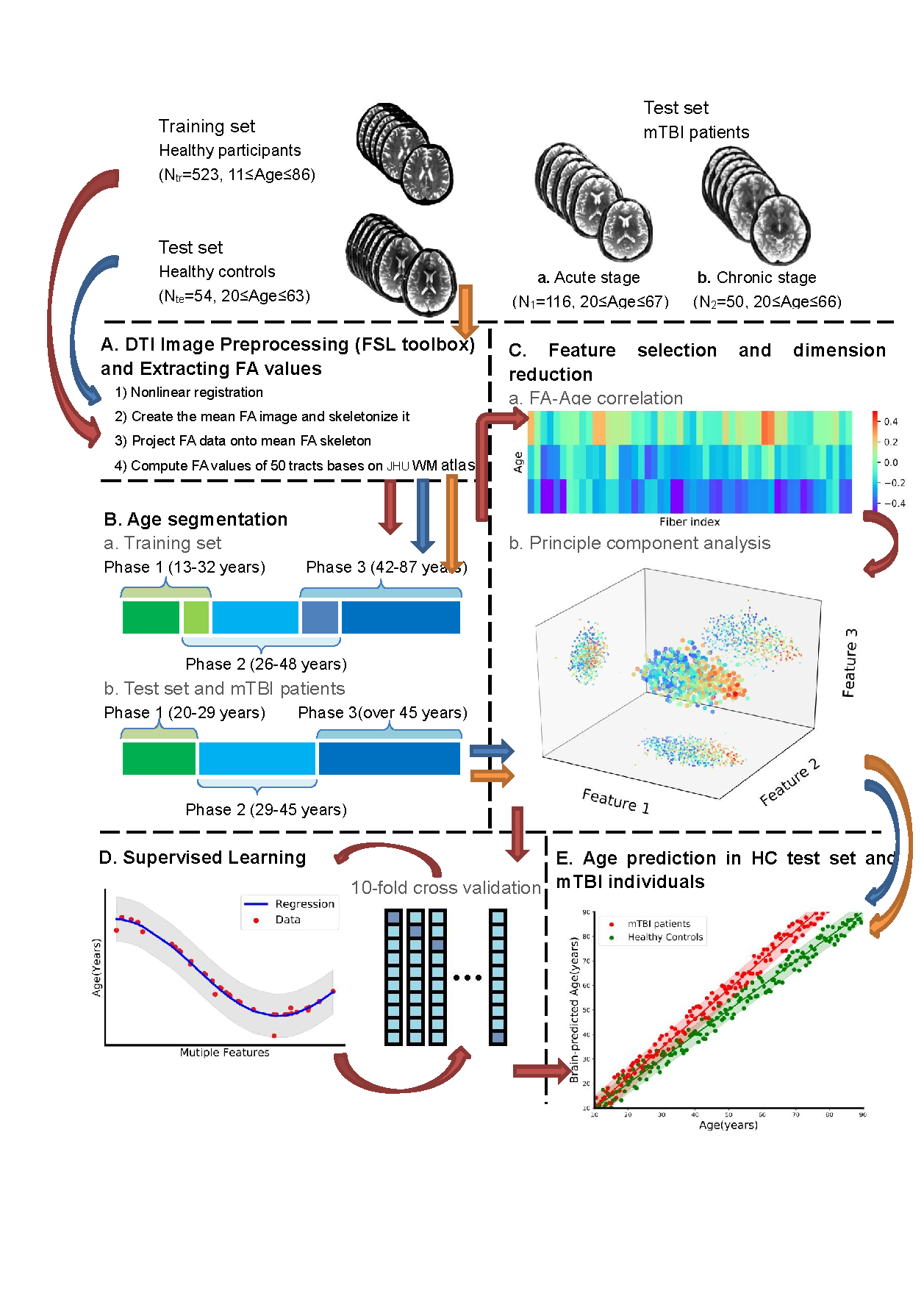
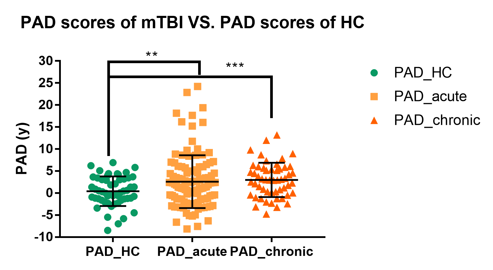
Figure 2 Comparing PAD scores of mTBI acute stage and chronic stage with HC. PAD scores of mTBI individuals at acute stage was significant greater than HC PAD scores (p < 0.01). PAD scores of mTBI individuals at chronic stage was significant greater than HC PAD scores (p < 0.001).
Note: *** denote p < 0.001; ** denote p < 0.01
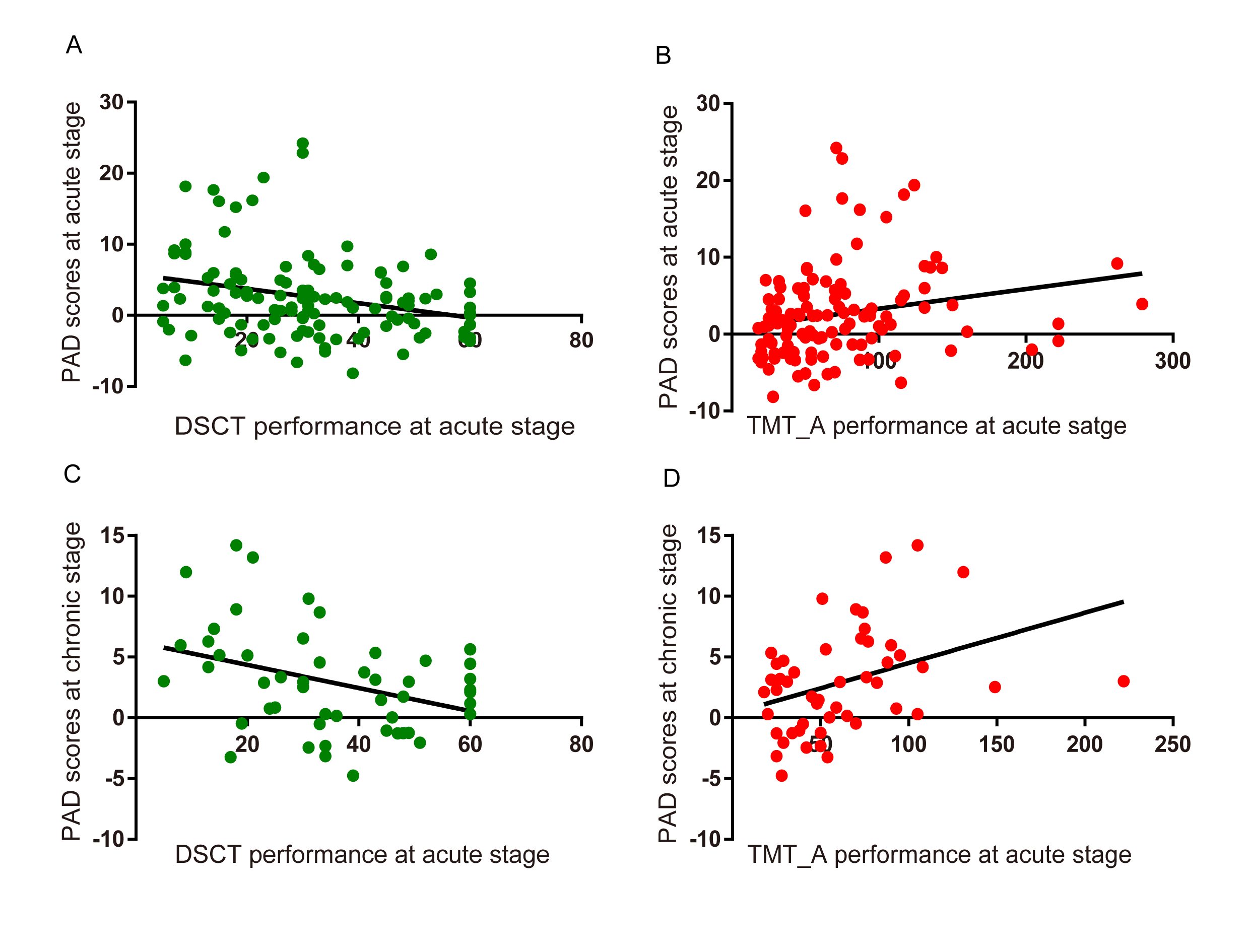
Figure 3. Information processing speed at acute stage predicts PAD scores across acute to chronic stage. A) Lower DSCT scores at acute stage significant correlated with PAD scores at acute stage (p < 0.01); B) Slower TMT_A performance at acute stage significant correlated with PAD scores at acute stage (p < 0.05); C) Lower DSCT scores at acute stage significantly predicted PAD scores at chronic stage (p < 0.01); D) Slower TMT_A performance at acute stage significantly predicted PAD scores at chronic stage (p < 0.01).
Note: DSCT = digital symbol coding test; TMT_A = trial making test A