0078
Free water elimination is necessary to characterize Diffuse Axonal Injury in moderate Traumatic Brain Injury1Penn Patho-Connectomics Lab, Radiology, University of Pennsylvania, Philadelphia, PA, United States, 2Molecular, Cellular, and Biomedical Sciences, CUNY School of Medicine, The City College of New York, New York, NY, United States
Synopsis
As the prevalence of diffusion MRI for clinical use grows, it is important to address the influence of injury-related extracellular water on the clinical interpretation of diffusion measures in conditions such as traumatic brain injury (TBI). The presence of extracellular free water from edema pollutes the estimation of diffusion measures, leading to flawed conclusions about the microstructure of the white matter. We demonstrate that Fernet, a robust single-shell free-water elimination method, can be used to decouple the effects of extracellular edema and tissue damage, to improve clinical understanding of the effects of injury on underlying white matter structure.
INTRODUCTION
Accurate estimation of measures obtained from diffusion MRI (dMRI) studies of moderate to severe Traumatic Brain Injury (TBI) is a clinically significant problem. The presence of increased water in the extracellular space of the brain contaminates the dMRI signal, resulting in ambiguity in interpretation of diffusion tensor (DT) measures1, and confounding clinical interpretations of white matter (WM) integrity in TBI patients. Free-water elimination (FWE) is an alternative to the common DT fit to dMRI data, which divides the signal into isotropic extracellular compartment representing free water (fw) and an anisotropic compartment representing tissue. Current state-of-the-art FWE methods require advanced multi-shell diffusion sequences, but single-shell acquisitions are still ubiquitous in the clinic. We propose the use of Fernet2, a robust single-shell FWE protocol shown to accurately estimate the fw compartment in simulated and human data, to aid in characterization of the effects of injury and recovery in WM in a longitudinal study of moderate to severe TBI patients. We hypothesize that Fernet enables interpretations axonal swelling and damage after TBI via its free-water and tissue compartments, that are ambiguous in a standard DT fit.METHODS
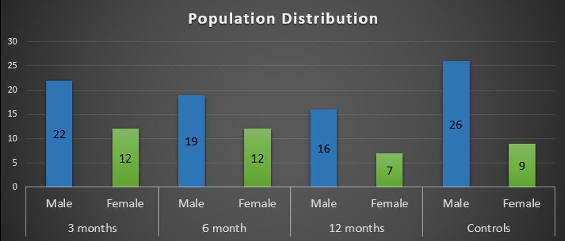
We used dMRI data of 45 moderate to severe TBI patients (with at most 3 scans at 3, 6, 12 months post-injury) and 35 healthy controls (Fig 1). dMRI was acquired at TR/TE=6,500/84 ms, 30 directions (b=1000s/mm2) and 7 b=0 images, repeated twice. Data was denoised, eddy-, motion- and bias-corrected. A standard tensor model (std) was fit to each voxel and diffusion measures of fractional anisotropy (FA-std) and axial diffusivity (AX-std) were computed. Fernet was used to generate free water (fw)-corrected diffusion measures FA-fw and AX-fw, and the free-water volume fraction (VF). Voxelwise t-tests (controlled for age and sex, corrected for multiple comparisons) on VF maps of patients against controls were computed at all three time points to elucidate the presence of extracellular water. Knowing that the presence of an isotropic compartment would affect the diffusion measures, we repeated the same t-tests on AX and FA maps, both ‘std’ and ‘fw’.RESULTS
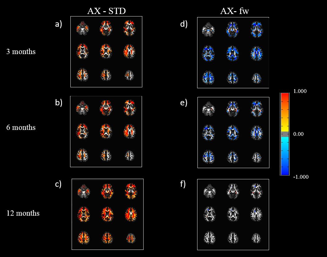
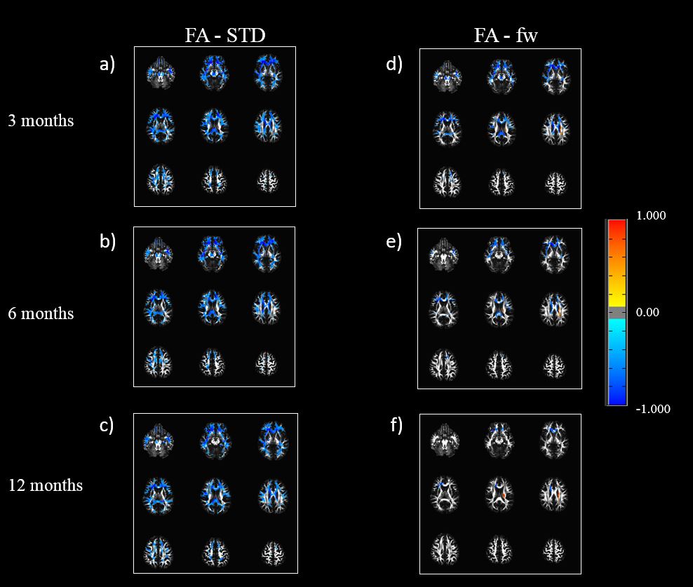
VF maps represent the isotropic compartment ‘removed’ through FWE. VF was significantly increased in patients at each time point (see Fig 2). Patients had a significantly higher AX-std than controls. The number of significant voxels increased over time with patients diverging from controls with time (Fig3 (a, b, c)). AX-fw maps showed a reduction in patients at the first time point that continued to increase with time, tending towards the controls’ baseline (Fig3 (d, e, f)). FA-std was lower in patients than controls, that continued to increase across time, tending to the controls’ baseline (Fig 4 (a,b,c)). FA-fw revealed a similar trend of decreased FA-fw in patients increasing across time, but the number of WM voxels showing a significant effect was lower than that of the FA-std maps (Fig 4 (d,e,f)).DISCUSSION
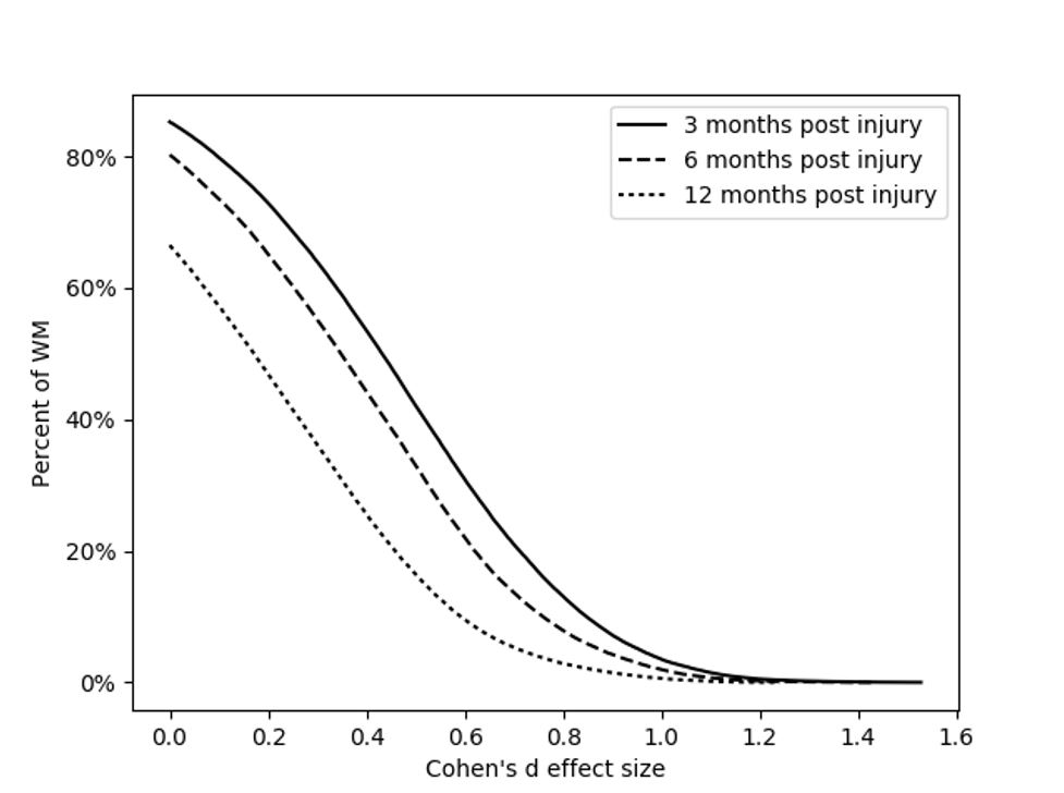
A major contributor to the neurocognitive deficits in TBI patients is Diffuse Axonal Injury (DAI)3, which results in widespread disruption in metabolic processes and architecture of axons. This eventually leads to a buildup in free water around axonal bodies, making the interpretation of diffusion measures challenging and unreliable. Axial Diffusivity reflects the underlying integrity and orientation of the WM fiber bundle. Thus, an increased AX-std implies an increased fiber integrity in patients, which is spurious as s DAI should manifest as a reduction in AX due to a breakdown in fiber integrity. The increased VF in the patients could be attributed to pathology related swelling and underlines the need for FWE. On doing so using Fernet, patients showed a lower AX-fw tending towards the controls’ baseline with time. VF is higher in patients and trends towards the controls as well. This suggests that the effect of DAI-induced reduced AX seen in AX-fw, was obscured in AX-std by the presence of extracellular water that drives diffusivity up. Fig 5 shows us that with each time point, the number of voxels with weaker effect sizes reduces, possibly denoting recovery in regions that were not as severely affected by the injury. The results in FA-std and FA-fw indicates the free-water compartment was artificially lowering the FA values in the FA-std maps, as well.CONCLUSION
Our results show that it is important to use free water elimination in TBI studies, in order to recover and report true underlying white matter microstructure. Additionally, free-water corrected AX and VF maps are more sensitive to TBI-induced changes in WM than FA. Thus, FWE paves the way for better TBI studies.Acknowledgements
This research was supported by the PA Department of Health award and the following National Institutes of Health (NIH) grants: 1R01NS096606 (PI: Ragini Verma) & R01NS092398 (PI: Junghoon John Kim).
References
1. Pasternak, O., Sochen, N., Gur, Y., Intrator, N., & Assaf, Y. (2009). Free water elimination and mapping from diffusion MRI. Magnetic Resonance in Medicine: An Official Journal of the International Society for Magnetic Resonance in Medicine, 62(3), 717-730.
2. Abdol Aziz Ould Ismail, Drew Parker, Moises Hernandez-Fernandez, Steven Brem, Simon Alexander, et al.. Characterizing Peritumoral Tissue Using Free Water Elimination in Clinical DTI. MICCAI 2018 - 21st International Conference on Medical Image Computing and Computer Assisted Intervention ; Workshop : Brain Lesion, Sep 2018, Granada, Spain. pp.1-9.
3. Dollé, J. P., Jaye, A., Anderson, S. A., Ahmadzadeh, H., Shenoy, V. B., & Smith, D. H. (2018). Newfound sex differences in axonal structure underlie differential outcomes from in vitro traumatic axonal injury. Experimental neurology, 300, 121-134.
Figures
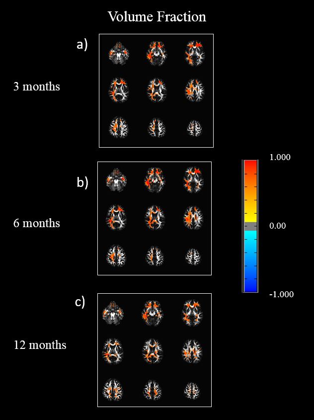
Results of the voxel-wise t-test, showing the volume fraction trend, across time. Figures (a), (b), and (c), show the t-test of volume fraction maps across three time points. All results are FDR corrected (q <0.0005)