0061
Beyond the Standard Model in Spinal Cord1Center of Functionally Integrative Neuroscience (CFIN) and MINDLab, Department of Clinical Medicine, Aarhus University, Aarhus, Denmark, 2Department of Physics and Astronomy, Aarhus University, Aarhus, Denmark, 3Champalimaud Neuroscience Programme, Lisbon, Portugal
Synopsis
A prevalent model for the diffusion signal from
Introduction
Resolving microstructure below nominal imaging resolution motivated the development of the so-called “Standard Model”[1] (SM), a compartment model of diffusion representing axons as “sticks”, i.e., with zero radial diffusivity. This invokes the assumption that the diffusion time is long enough for the intra-axonal radial diffusivity to have reached its asymptotic limit of zero[1]. This may be justified in the brain where axons are typically very small; however, simulations have recently shown that spinal cord diameters may be sufficiently large to invalidate this assumption up to large diffusion times[2]. Here, we extend the SM by adding an intra-axonal radial diffusivity. Using diffusion data from a rat spinal cord and thereon based simulations, it is shown that reliable model parameter estimation is feasible given an extensive dataset. Furthermore, applying the extended model to the spinal cord data leads to unphysical parameter estimates revealing the inapplicability of the SM in this tissue.Methods
The considered version of the SM consists of an intra- and extra-axonal compartment as detailed in [3] with signal expression
$$S(b,\boldsymbol{\hat{g}}) = \int d\boldsymbol{\hat{n}} \mathcal{P}(\boldsymbol{\hat{n}}) \left( fe^{-bD_a(\boldsymbol{\hat{g}}\cdot\boldsymbol{\hat{n}})^2} + (1-f)e^{-bD_e^\perp-b(D_e^\parallel-D_e^\perp)(\boldsymbol{\hat{g}}\cdot\boldsymbol{\hat{n}})^2} \right)$$
where $$$\mathcal{P}$$$ is the fibre ODF, $$$f$$$ the intra-axonal volume fraction, $$$D_a$$$ the intra-axonal longitudinal diffusivity, and $$$D_e^\parallel$$$ and $$$D_e^\perp$$$ are respectively the extra-axonal longitudinal and radial diffusivities. Extending with an intra-axonal radial diffusivity $$$D_a^\perp$$$ simply entails introducing it in the intra-axonal expression identically to how $$$D_e^\perp$$$ appears in the extra-axonal one.
Model parameters are estimated by non-linear fitting of the model-representation in spherical harmonics (fibre ODF) and Legendre polynomials (kernel) up to order $$$l=8$$$[4]. For fit initialization, parameters are estimated by matching the model’s moments to cumulant estimates obtained from an axisymmetric DKI fit[5] to the subset of data with b$$$\leq$$$3ms/μm2 as detailed in[3] but without assuming an ODF. Consequently, the SM parameters are parametrized by the $$$l=2$$$ ODF Legendre expansion coefficient. A discrete subset of initializations is utilized by varying the expansion coefficient in discrete steps. Therefore, each signal realization results in multiple candidate parameter estimates – many of which are equal or very similar. When fitting the extended model, $$$D_a^\perp$$$ is initialized to zero.
Spinal Cord Data
All experiments were preapproved by the local animal ethics committee operating under local and EU laws. The rat spinal cord was extracted as previously described[6]. A 16.4T Bruker Aeon Ascend magnet with a 5mm birdcage coil mounted on a micro5 probe capable of producing up to 3000mT/m isotropically was employed. The spinal cord was placed in a Fluorinert-filled 5mm NMR tube and kept at 37C throughout the experiments. Single Diffusion Encoding (SDE)[7] data was recorded using an EPI readout [FOV: 6x6mm2, acquisition matrix: 70x70, in-plane resolution: 86x86μm2, slice thickness: 1.35mm]. The number of gradient directions was 64 in each of 33 b-shells linearly varied from 0 to 9ms/μm2. Δ/δ=45/2ms. Data was denoised[8] and corrected for Rician bias[9] and Gibbs ringing[10] prior to further analysis.
Only WM voxels identified by having FA>0.7 (fig. 1).
Results
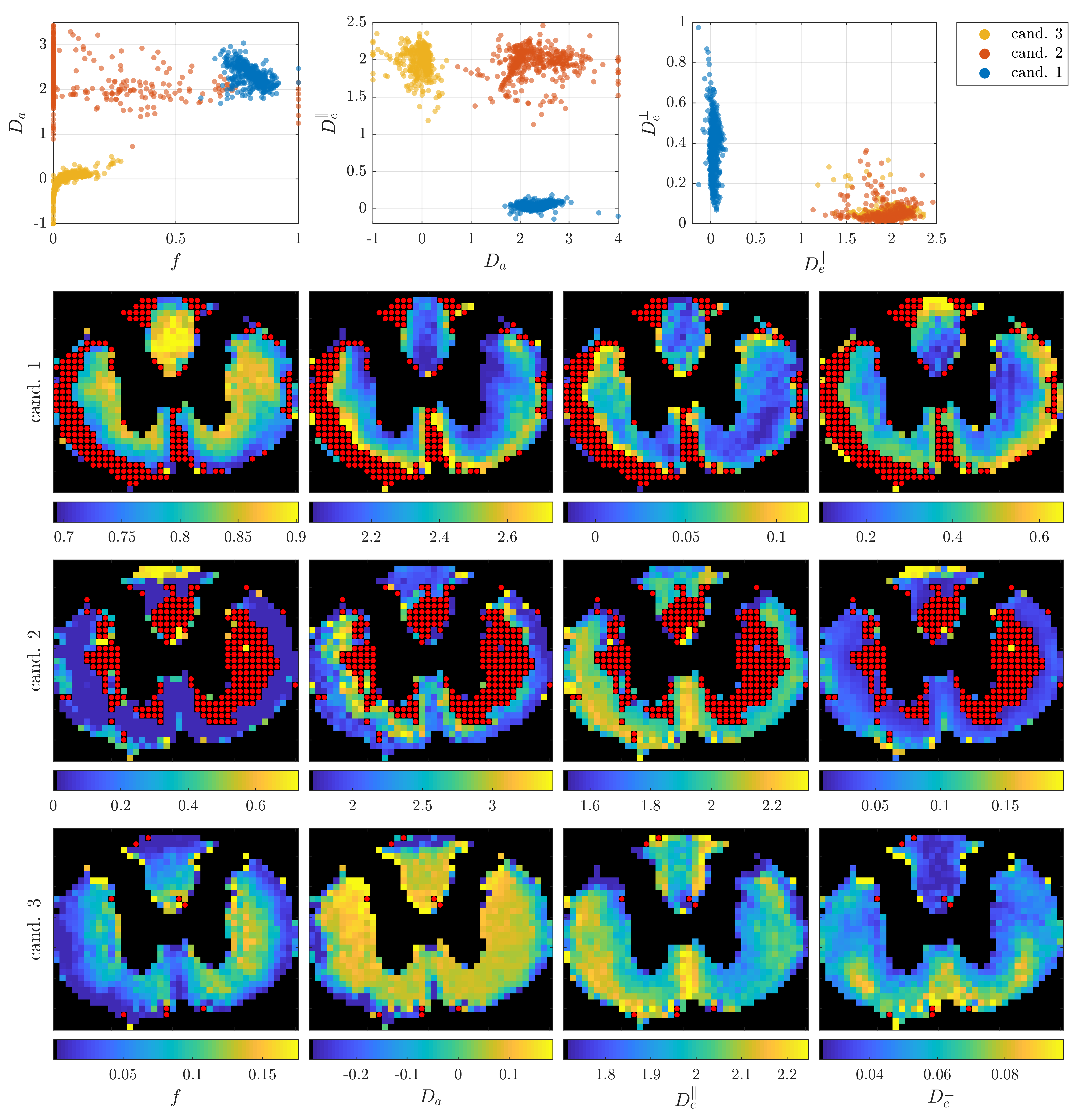
Initially, parameters of the non-extended SM were estimated. This results in three groups of parameter candidates shown in fig. 2: candidate 2 is disregarded for having low fitting quality and mostly identifying $$$f=0$$$ resulting in an implausible one-compartment model. Candidate 3 is found to have the best fitting quality in 95% of voxels suggesting its physical plausibility, with $$$D_a<D_e^\parallel$$$ in contradiction to previous studies[3,11] and additionally proposes a very small axonal volume fraction.
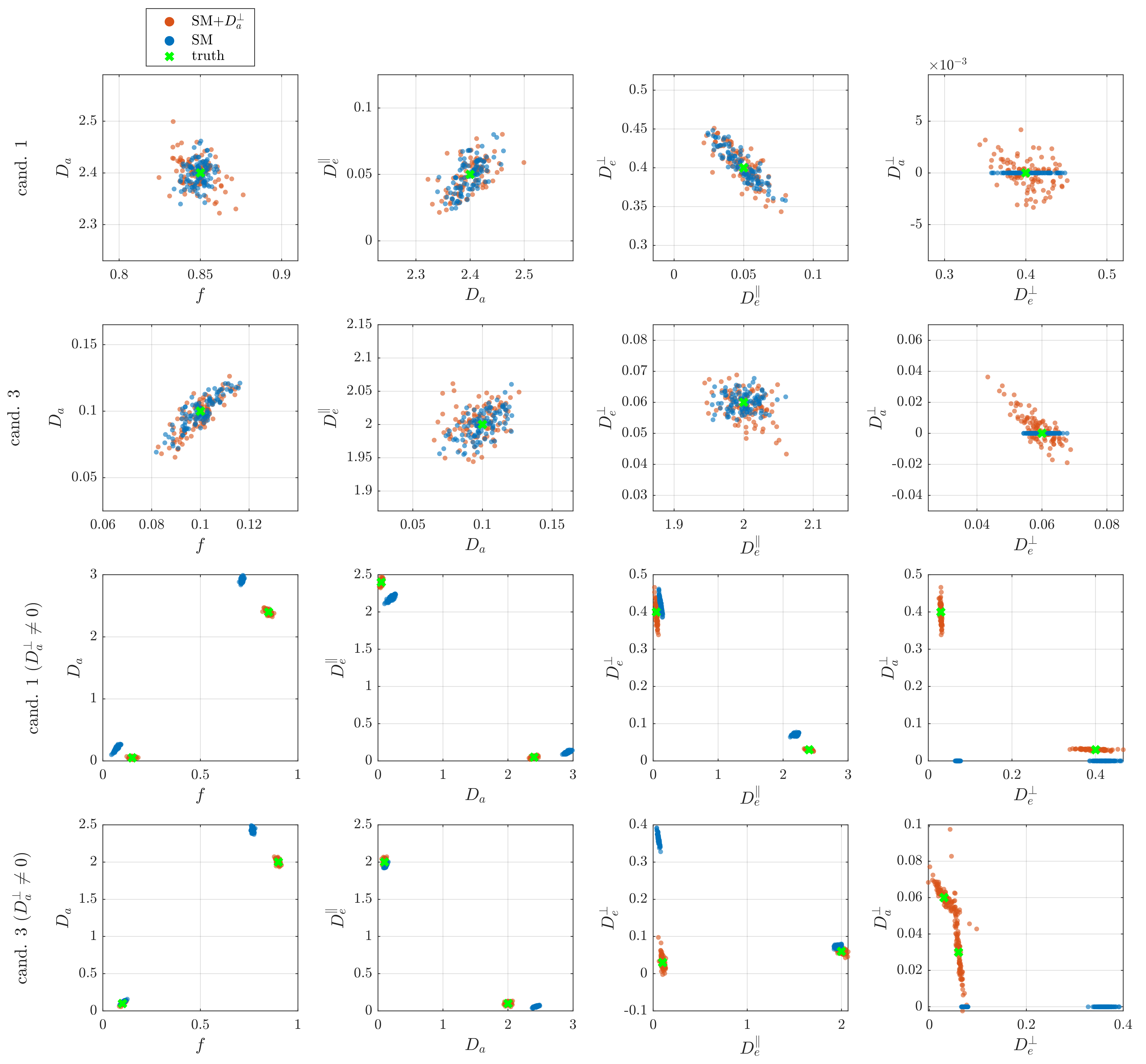
Simulations with an a-priori known ground truth were used to assess the reliability of the parameter estimates. The results in fig. 2 indicate that with the present extensive dataset, estimates are reliable assuming that the SM does model the tissue well. Furthermore, it is indeed feasible to extend with $$$D_a^\perp$$$ which will be estimated to zero if negligible. Interestingly, it is found that even small values of $$$D_a^\perp$$$ (~0.03) can cause considerable bias if ignored.
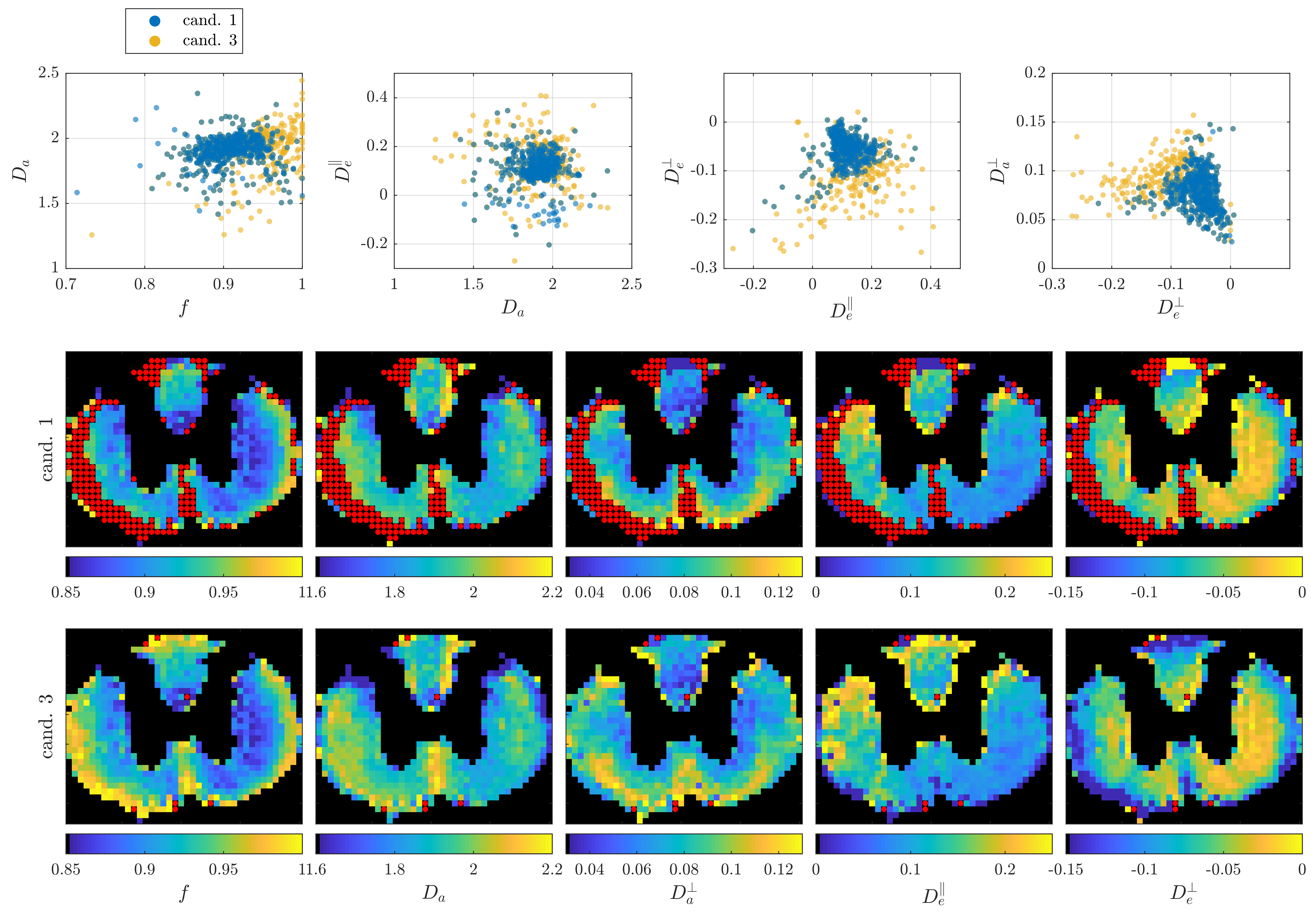
Fitting the extended model to the rat SC data (fig. 4) results in the same parameter estimates for groups 1 and 3 reducing the number of candidates to one. Intriguingly, the radial diffusivity in one compartment is estimated to be negative thereby indicating that the two-compartment SM is insufficient to model this data.
Conclusion
Using an extensive diffusion MRI dataset of a rat cervical spinal cord, estimated SM parameters with the extension of an intra-axonal radial diffusivity were presented. Based on simulations these were shown to be reliable given that the model applies. The estimate being unphysical is therefore taken as an indication that a two-compartment SM is insufficient to model the data.Acknowledgements
No acknowledgement found.References
[1] D. S. Novikov et. al.: “Quantifying brain microstructure with diffusion MRI: Theory and parameter estimation”, NMR in Biomedicine (2018).
[2] F. Grussu et. al: “Relevance of time‐dependence for clinically viable diffusion imaging of the spinal cord”, Magnetic Resonance in Medicine (2018)
[3] S. N. Jespersen et. al.: “Diffusion time dependence of microstructural parameters in fixed spinal cord”, NeuroImage 182 (2018)
[4] S. N. Jespersen et. al.: “Modeling dendrite density from magnetic resonance diffusion measurements”, NeuroImage 34 (2007)
[5] B. Hansen et. al: “Fast imaging of mean, axial and radial diffusion kurtosis”, NeuroImage 142 (2016)
[6] D. Nunes et. al.: “Mapping axonal density and average diameter using non-monotonic time-dependent gradient-echo MRI”, Journal of Magnetic Resonance 277 (2017)
[7] N. Shemesh et. al.: “Conventions and nomenclature for double diffusion encoding NMR and MRI”, Magnetic Resonance in Medicine 75 (2016)
[8] J. Veraart et. al.: “Denoising of diffusion MRI using random matrix theory”, NeuroImage 142 (2016)
[9] C. G. Koay et. al.: “Analytically exact correction scheme for signal extraction from noisy magnitude MR signals”, Journal of Magnetic Resonance 179 (2006)
[10] E. Kellner et. al.: “Gibbs-ringing artifact removal based on local subvoxel-shifts”, Magnetic Resonance in Medicine 76 (2016)
[11] N. Kunz et. al.: “Intra- and extra-axonal axial diffusivities in the white matter: Which one is faster?”, NeuroImage 181 (2018)
Figures