0055
Identifying focal thalamic activity underlying sleep and wake states through EEG-fMRI at 7 Tesla1Boston University, Boston, MA, United States, 2Massachusetts General Hospital, Boston, MA, United States, 3Massachusetts General Hospital/Harvard Medical School, Boston, MA, United States, 4BIDMC/Harvard Medical School, Boston, MA, United States
Synopsis
The thalamus plays an important role in regulating brain states, but remains poorly understood due to the technical challenges in imaging small brain structures with simultaneous electrophysiology. We implemented simultaneous fast fMRI and EEG at 7 Tesla to achieve high-SNR imaging of thalamic dynamics during human sleep. We found that we could detect selective activity within a focal set of thalamic nuclei that preceded the moment of awakening. These results identify potential network mechanisms engaged in regulating brain states, and demonstrate the potential for multimodal 7T imaging to identify new roles for deep brain structures in regulating cortical function and cognition.
Introduction
Brain arousal states are dynamically modulated throughout the day, and can shift within hundreds of milliseconds – for example, at the transition between sleep and wake. Animal studies have suggested an important role for the thalamus in controlling brain states and cortical dynamics, but human studies have been limited, due to the technological challenges in measuring activity in small, deep brain structures in humans. EEG tracks fast changes in cortical dynamics, but cannot detect neural activity in deep structures such as the thalamus. Furthermore, thalamus is made up of functionally diverse nuclei, and the roles of these substructures in modulating brain states remains poorly understood. Ultra-high field fMRI could enable detection of activity within these small thalamic nuclei, but recording EEG at 7 Tesla is technically challenging due to the severe artifacts induced by the static magnetic field. We developed a simultaneous EEG and fast fMRI paradigm at 7 Tesla to track dynamics within individual thalamic nuclei, and examined their relationship to cortical electrophysiology and arousal.Methods
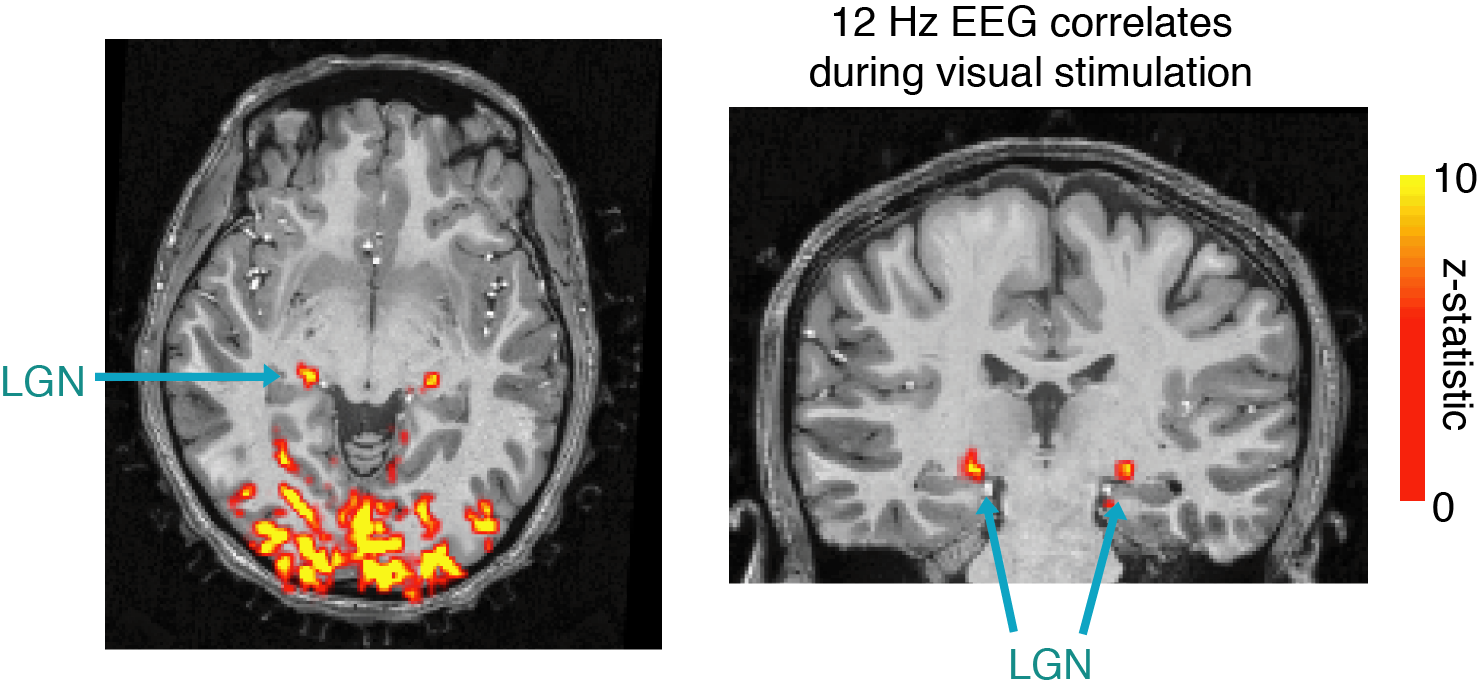
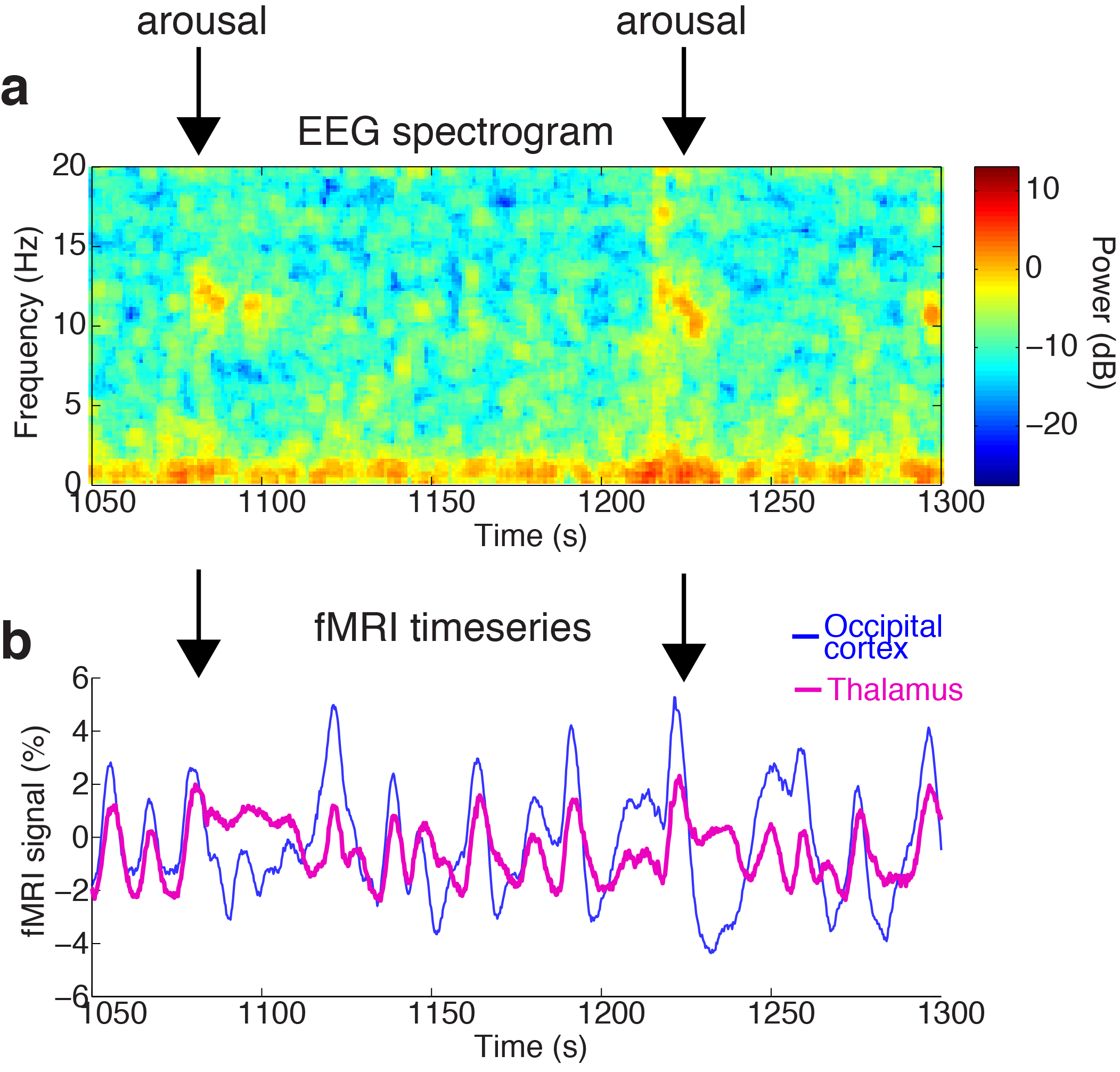
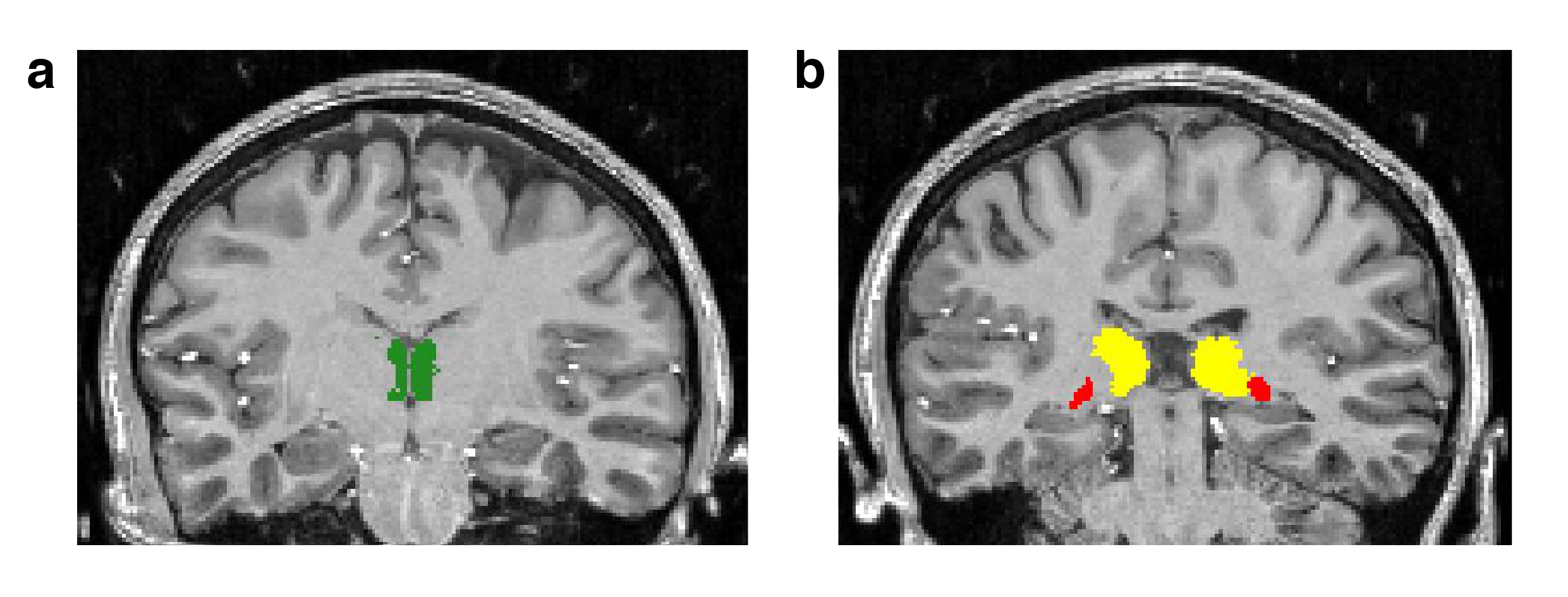
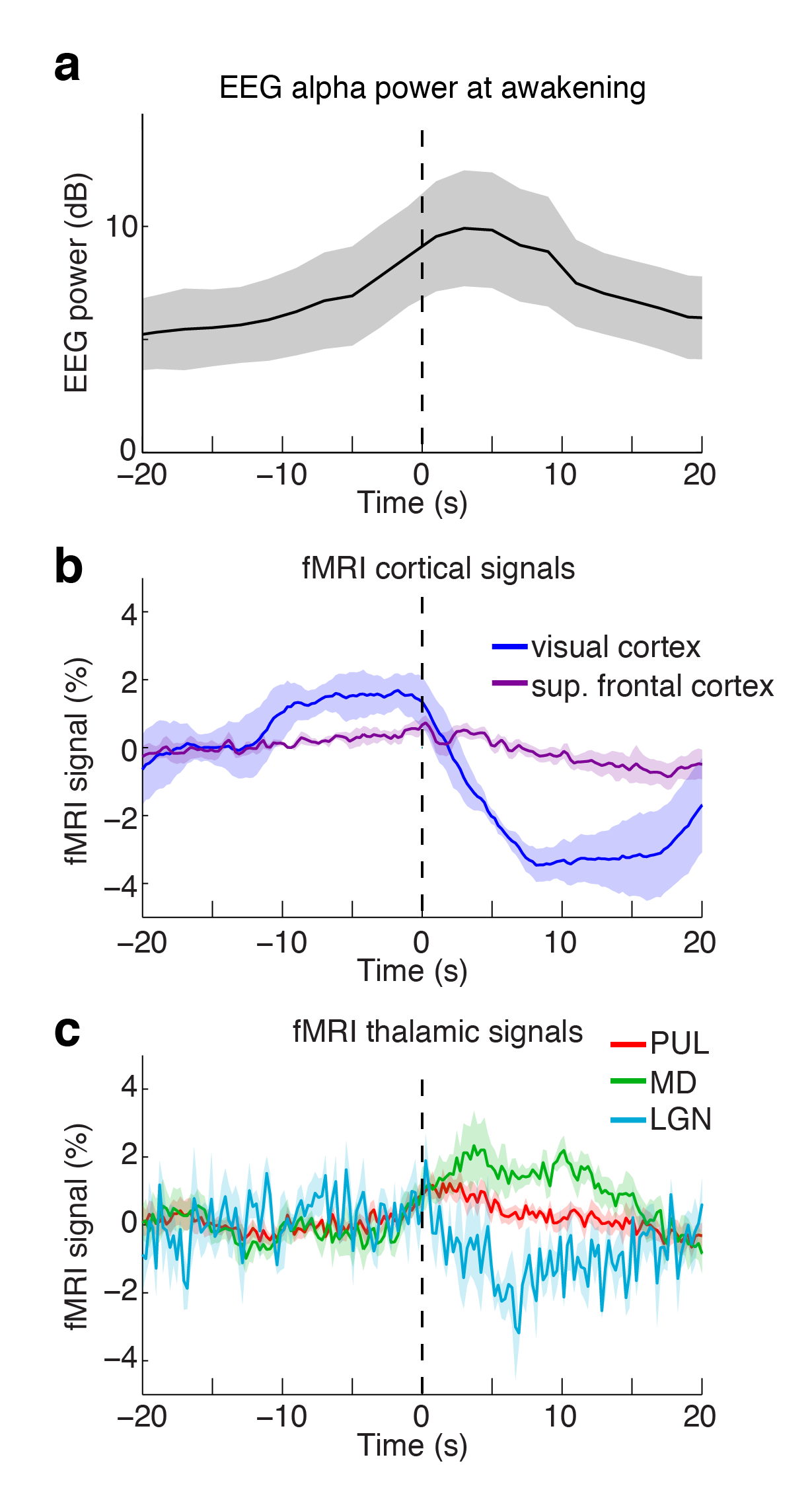
Five subjects gave informed consent and were scanned on a 7T Siemens whole-body scanner with a custom 32-channel head coil, as they slept inside the scanner for up to two hours. EEG was simultaneously acquired using a polymer thick film-based InkNet designed for use at 7T 1. Artifacts in the EEG signal were removed using a time-varying reference-based cleaning approach adapted from prior methods 2. Sessions began with a 750-micron resolution T1-MEMPRAGE, and then resting-state BOLD fMRI data were acquired with a single-shot gradient echo (TR=247 ms, SMS=8, 2.5 mm isotropic resolution). Data were motion corrected, slice timing corrected, and physiological noise was removed through a sliding window adaptation of RETROICOR to accommodate long duration scans. For the visual validation, 12 Hz EEG power was extracted and convolved with a standard hemodynamic response function. Statistical maps were computed in FSL. For the sleep study, three thalamic nuclei with relatively large volumes and were automatically segmented from the T1 anatomical data 3: lateral geniculate nucleus (LGN, primary visual); pulvinar (PUL, higher-order visual); mediodorsal (MD, higher-order frontal attentional). Spontaneous awakenings were identified manually using the alpha (~10 Hz) power of the EEG data, and then the mean signal in each nucleus was computed, locked to the time of awakening.Results
We first validated that our 7T EEG-fMRI approach yielded sufficiently high-quality data to resolve EEG oscillations and local thalamic fMRI activity, using a visual experiment. We correlated the fMRI signal with the envelope of the stimulus-induced 12 Hz EEG flicker (convolved with an HRF), and observed clear activation both throughout visual cortex, and specifically within the LGN of the thalamus (Fig. 1).
We next analyzed fMRI dynamics around the time of spontaneous awakening from sleep, defined through the change in cortical electrophysiology detected in the EEG (Fig. 2a). We observed clear thalamic fMRI activations at each arousal from sleep (Fig. 2b). To determine whether distinct dynamics in thalamic nuclei were associated with this transition, we examined the mean timeseries in ROIs relative to the moment of awakening (Fig. 3). Awakening was not associated with a strong increase in cortical activity (Fig. 4a,b). However, we observed distinct increases in selective activity within the MD and PUL thalamic nuclei upon awakening. In contrast, LGN activity was suppressed after awakening from sleep, potentially reflecting that the eyes remained closed, while sleep-related activity ceased. The timescale of the MD activation showed an increase within a second of awakening – due to the ~4-5 second hemodynamic lag of fMRI signals, this suggested that these focal thalamic activations preceded the change in cortical electrophysiological state.
Discussion
We find that 7T fast fMRI-EEG detects increased activity within individual thalamic nuclei that occurs immediately prior to awakening from sleep. Previous studies have shown that deactivations throughout the thalamus during low arousal states 4; our work is consistent with these results, and further demonstrates that within the thalamus, specific subnuclei exhibit distinct dynamics locked to the transitions between arousal states. These results suggest that the mediodorsal nucleus may be selectively engaged at the transition between sleep and wake, and could potentially drive subsequent changes in cortical oscillatory signals. Our results also demonstrate the potential for EEG-fMRI at 7T to dissect the large-scale network mechanisms that underlie regulation of cortical dynamics and brain state.Acknowledgements
This work was supported in part by NIH grants K99-MH111748, R01-EB019437, S10-RR023043 and S10-RR019371 , and by the MGH/HST Athinoula A. Martinos Center for Biomedical Imaging.References
1. Poulsen, C. et al. Polymer thick film technology for improved simultaneous dEEG/MRI recording: Safety and MRI data quality. Magn. Reson. Med. 77, 895–903 (2017).
2. Luo, Q., Huang, X. & Glover, G. H. Ballistocardiogram artifact removal with a reference layer and standard EEG cap. Journal of Neuroscience Methods 233, 137–149 (2014).
3. Iglesias, J. E. et al. A probabilistic atlas of the human thalamic nuclei combining ex vivo MRI and histology. Neuroimage 183, 314–326 (2018).
4. Chang, C. et al. Tracking brain arousal fluctuations with fMRI. Proc Natl Acad Sci USA 113, 4518–4523 (2016).
Figures