0046
Novel methods to record MR spectra in human brain without suppressing or exciting the water signal to investigate exchange-sensitive protons.1Departments of Radiology and Biomedical Research, University Bern, Bern, Switzerland, 2Department of Biomedical Imaging and Image-guided Therapy, Medical University Vienna, High-field MR Center, Vienna, Austria
Synopsis
Non-water-suppressed MR spectroscopy in the form of metabolite-cycled MRS and longitudinal-relaxation-enhanced MRS (also termed non-water-excitation, NWE) has come into focus when studying compounds with exchanging protons (observed in the downfield region) or compounds like NAD+ that seem to be affected by cross relaxation from water. Here, we strive to implement new NWE techniques that can be used at 3T and allow for very short TE and observation of fast exchanging protons. 2D I-CSE, a combination of 2D ISIS and slice-selection with chemical shift selective excitation, fulfills this profile and first human brain applications show large signal contributions from exchanging protons.
Introduction
Most clinical MR spectroscopy studies focus on the upfield part of the spectrum, while the downfield region is neglected. Obvious reasons are the low SNR and the fact that downfield peaks are influenced by exchange effects. Previous research includes determination of exchange constants at 3T and at ultra-high fields, using techniques that aim to show minimal influence from water saturation – in particular using the metabolite-cycling (MC) technique1 where the effective time for magnetization exchange between water and metabolite signal is usually >20ms. Alternatively, implemented only for ultra-high field, non-water-excitation (NWE) techniques2 rely on leaving longitudinal water magnetization undisturbed and make use of chemical-shift-selective excitation in combination with LASER localization.
Here, we strive to implement new NWE techniques that can be used at 3T and allow for very short TE and observation of fast exchanging protons.
Methods
The first proposed technique (Fig. 1a), termed 3D I-CSE, uses ISIS localization with slice-selective adiabatic GOIA-W(16, 4)3 inversion pulses and the excitation performed with a Chemical-shift-Selective Excitation module. In the second sequence, 2D I-CSE (Fig. 1b), one ISIS localization pulse is replaced by a slice-selective refocusing pulse. Chemical-shift-selective excitation (and refocusing in 3D I-CSE) uses Gaussian-modulated frequency-selective pulses. For slice-selection in 2D I-CSE a Mao refocusing pulse is applied. Both sequences are preceded by full outer-volume suppression4. Minimal TE depends on the excitation pulse and was <10ms in both presented cases. An additional water inversion pulse was implemented to precede the ISIS module. It can be activated for those scans in the ISIS-cycle that leave the water magnetization in the ROI inverted. This will allow longitudinal relaxation-enhancement (LRE)2 in every acquisition. Water pre-saturation (WS) by CHESS was added to investigate saturation-transfer effects from water.
Measurement parameters: spectral width 2500Hz, 128 averages, TR 3000ms, TE 8.4ms. Sequence tested on a ”braino” phantom. Five healthy volunteers examined on a 3T Siemens Prisma scanner using a multi-channel receive head coil. Data from a large ROI of 50x65x20 mm3 placed above the ventricles in two volunteers is presented to compare in vivo results between different sequences, where a semiLaser sequence with MC1 was used as another sequence without WS (TE 40ms).
Data processing done in jmrui and matlab. To correct for water sidebands (well visible in case of residual water excitation) NWE spectra were eddy-current corrected using a water reference acquired with on-resonance excitation.
Results & Discussion
MR spectra acquired with PRESS and the developed techniques in “braino” are presented in Fig. 2 and show very similar profiles, but also indicate the limited spectral range with full intensity in the I-CSE methods. As presented in Fig. 3, the excitation profile for 3D I-CSE is rather narrow and does not yield uniform excitation over an appreciable spectral range–though this could easily be optimized with adapted rf pulses. In contrast, 2D I-CSE offers a fairly uniform profile with small excitation >500Hz off-resonance. 2D I-CSE also has the advantage over the 3D ISIS variant that it does not rely on outer-volume signal cancelation over the whole head, but only the selected slice. This is illustrated in Fig.4, where single-shot acquisitions and the resulting signal from the ROI are shown.
Figure 5 presents the downfield spectra acquired for 2 volunteers using 3D I-CSE, 2D I-CSE, and MC-semiLaser, each with and without WS. The spectra demonstrate the advantage of 2D I-CSE for detection of exchangeable protons with negligible effect of water excitation and very short TE. The difference spectra clearly show large peaks for exchangeable amide protons >8 ppm and a clear exchange effect at 6.8ppm, while non-exchangeable resonances are cancelled proving the robustness of the technique. The semiLaser spectra also demonstrate some effect of WS, but the majority of signal from labile protons is suppressed/decayed in scans without WS because of long TE and the MC preparation period. MC combined with short-TE STEAM has shown stronger effects of WS1. However, the present technique certainly excels with better SNR and promises extension to detect faster exchanging compounds. The spectra in Fig. 5a acquired with the 3D I-CSE technique are of lower quality demonstrating some effect of exchange, but signal cancellation for localization based on a full ISIS scheme does not seem to work reliably.
Conclusions
The designed 2D I-CSE sequence offers non-water-suppressed and NWE acquisition of downfield spectra combined with an ultrashort TE at 3T – easily extendable to higher fields. It offers clear benefits compared to MC for observation of fast exchanging or relaxing compounds. Further work will aim at optimizing the selective excitation, improved phase-cycling, and at optimal detection of NAD+5 at clinical field strengths.Acknowledgements
This work is supported by the Swiss National Science Foundation (SNSF #320030‐175984).References
[1] Giapitzakis, et al. Metabolite-cycled STEAM and semi-LASER localization for MR spectroscopy of the human brain at 9.4T. Magn Reson Med 79:1841-1850, 2018.
[2] Shemesh, et al. Metabolic properties in stroked rats revealed by relaxation-enhanced magnetic resonance spectroscopy at ultrahigh fields, Nature Communications vol.5 4958, 2014.
[3] Bogner, et al. In Vivo 31P spectroscopy by fully adiabatic extended image selected in vivo spectroscopy: A comparison between 3 T and 7 T, Magn Reson Med 66:923–930, 2011.
[4] Connelly, et al. Outer volume suppressed image related in vivo spectroscopy (OSIRIS), a high-sensitivity localization technique. J.Magn.Reson. 78:519-525, 1988.
[5] de Graaf, et al. Detection of cerebral NAD+ in humans at 7T. Magn Reson Med 78:828-835, 2017
Figures
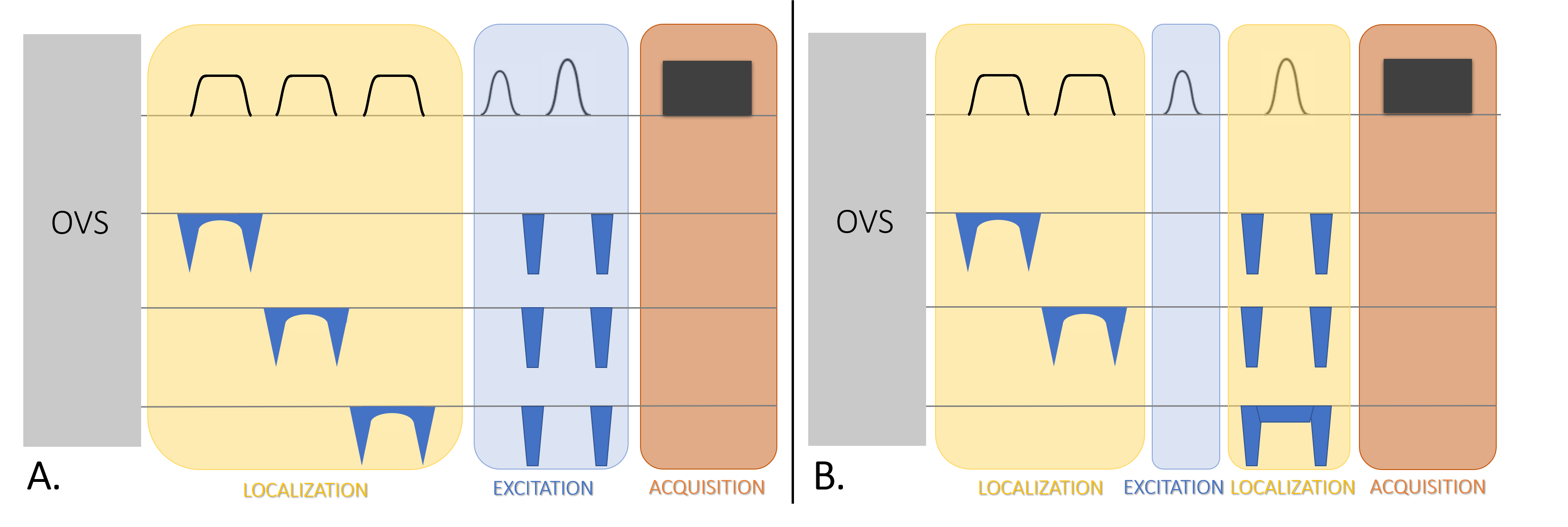
Figure 1. Schematic sequence diagram of the proposed non-water-excitation sequences. Left: Three-dimensional ISIS localization with Chemical-shift-Selective Excitation termed 3D I-CSE. Right: Two-dimensional ISIS localization with Chemical-shift-Selective Excitation and slice-selective spin echo, termed 2D I-CSE. In both sequences ISIS makes use of GOIA pulses and selective excitation is currently using Gaussian modulated pulses. Optionally, the ISIS module can be preceded by a further selective water inversion pulse in odd acquisitions to re-establish the longitudinal water magnetization in the region of interest along B0.
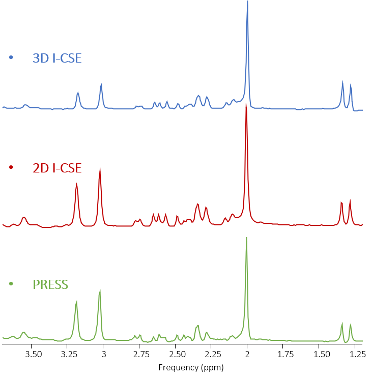
Figure 2. 1H MRS spectra comparing upfield spectra from a “braino” phantom as obtained with 3D I-CSE, 2D I-CSE and PRESS illustrating that both novel sequences perform well in the simple situation of a spherical phantom. The spectra were acquired with a frequency offset of -4ppm for the selective pulses( with respect to the water peak).
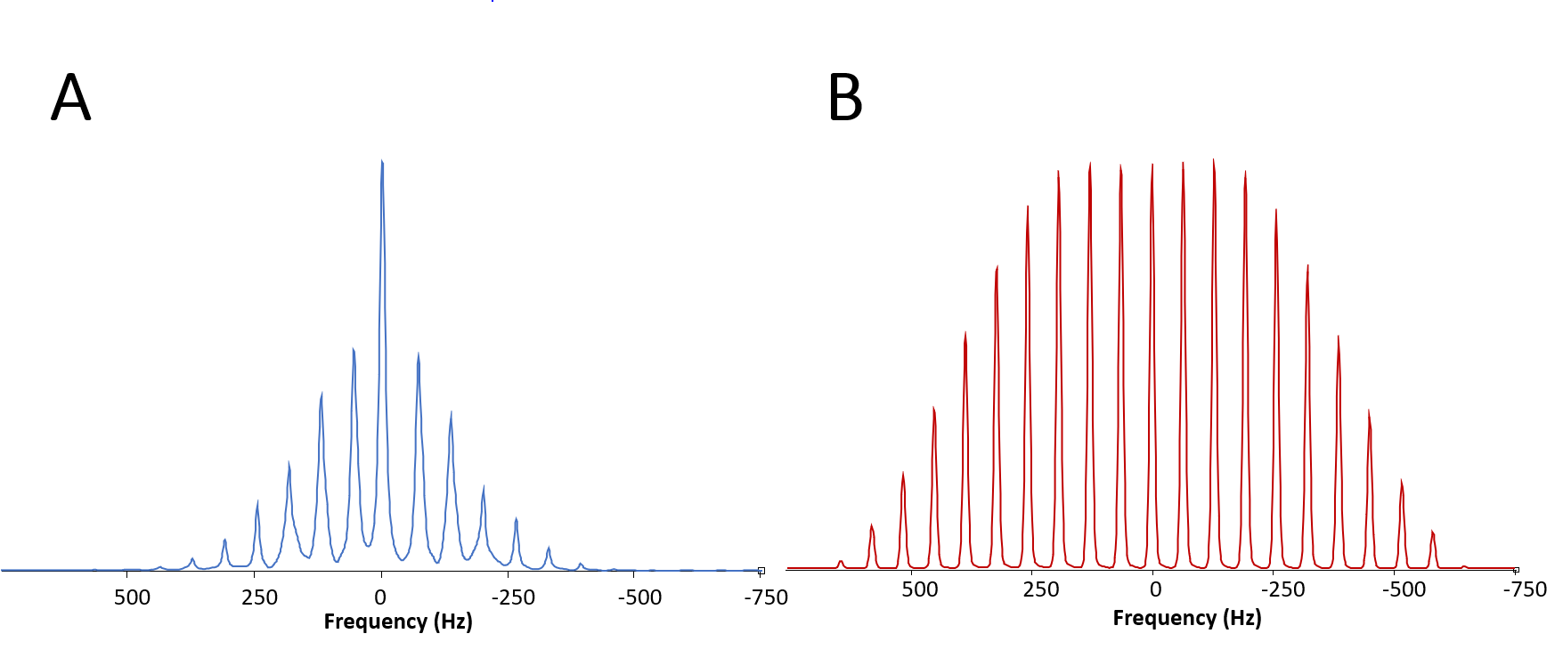
Figure 3. Excitation module profile for 3D I-CSE (A) and 2D I-CSE (B). The presented profiles were obtained from 21 measurements with varying frequency offset (from -5.0 ppm to 5.0 ppm with respect to the water peak in 0.5 ppm steps). The spectra were overlaid and broaden in jMRUI.
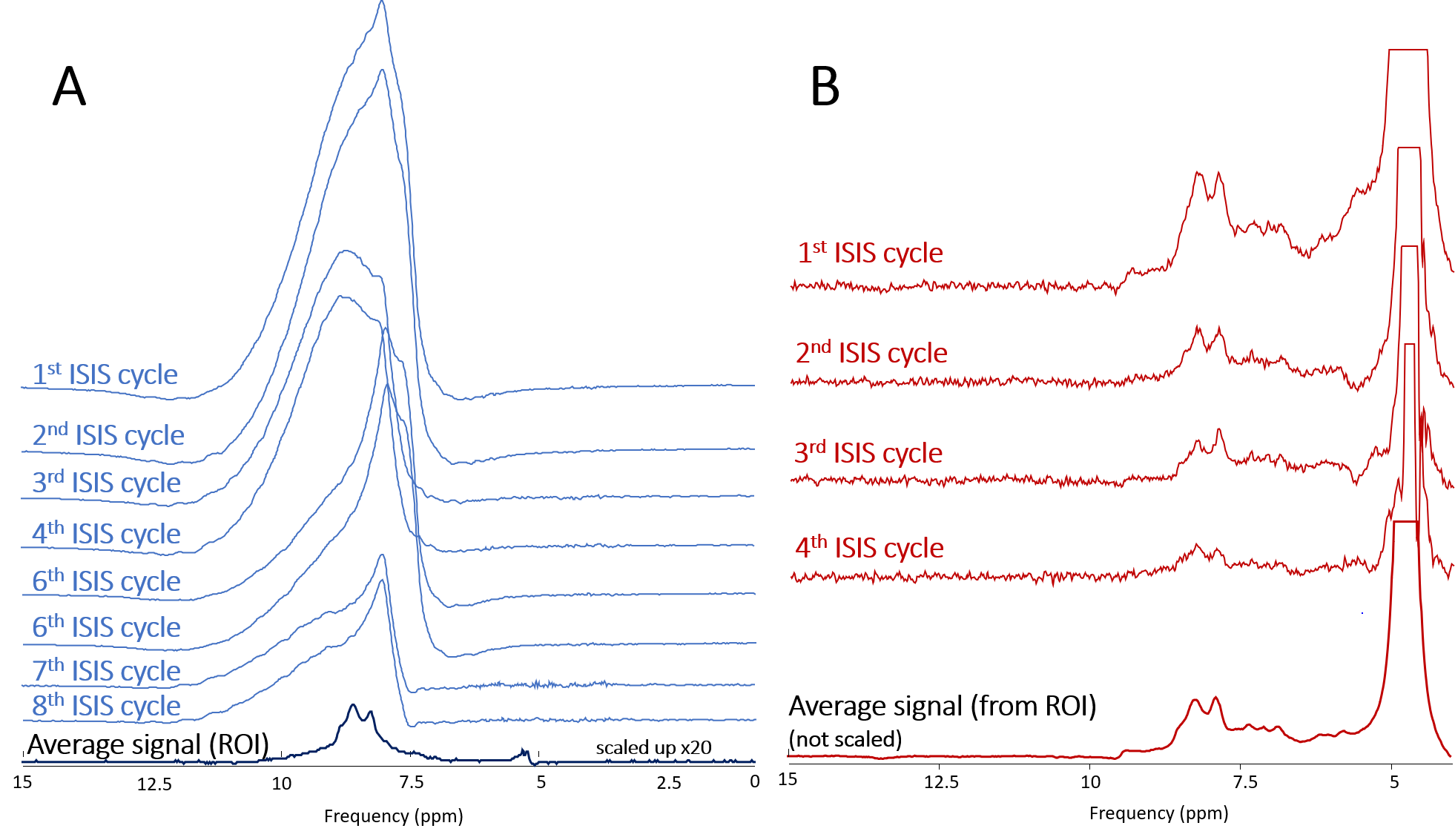
Figure 4. Illustration of single acquisitions and resulting average for 3D I-CSE (A) and 2D I-CSE (B). For the 3D ISIS technique the signal in each shot is a superposition from signal of the whole head, which is appreciated when comparing the size of single scans and the resulting signal from the ROI. Without instabilities, the outer-volume-signals cancel. (Single-shot signals phased to positive signal in this plot irrespective of ISIS cycle phase). The 2D I-CSE method clearly does not have a similarly sized problem with outer volume signal cancellation since only signal from one slice is recorded in each acquisition.
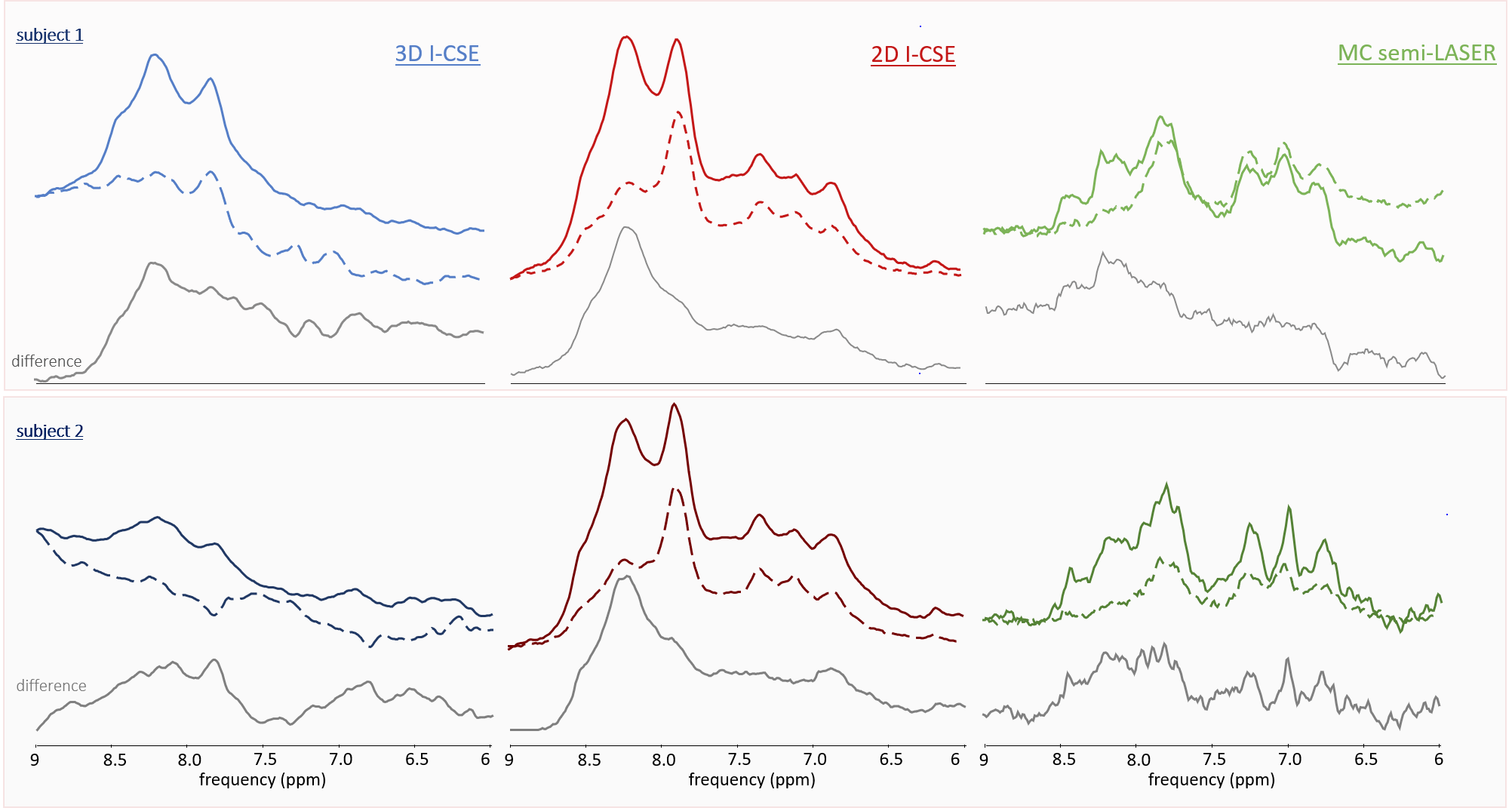
Figure 5. Comparison of in-vivo brain spectra from two healthy subjects acquired for the downfield region with 3D I-CSE (first), 2D I-CSE (second) and MC-semiLASER (third column) with (middle, dashed lines) and without (top, solid lines) water suppression. Difference spectra between water-suppressed and non-suppressed scans, plotted at the bottom, reflect signal suppressed via saturation-transfer by the CHESS water-suppression module. 3D I-CSE apparently failed for the second volunteer – possibly due to motion or system instability. (Excitation at 9.7ppm in I-CSI methods; difference spectra obtained after 3Hz Gaussian line-broadening and manual frequency alignment at the downfield NAA peak at 7.8ppm).