0045
Investigating the effects of an early intervention in preterm newborns: A resting-state functional connectivity study1Division of Development and Growth, Department of Pediatrics, University of Geneva, Geneva, Switzerland, 2Institute of Bioengineering, Ecole Polytechnique Federale de Lausanne (EPFL), Lausanne, Switzerland, 3Institute of Mathematics, Ecole Polytechnique Federale de Lausanne (EPFL), Lausanne, Switzerland
Synopsis
In this
Introduction
Recent advances in the Neonatal Intensive Care Unit (NICU), where preterm infants spend their first weeks of life, have led to an increase in the survival rate of these neonates. However, premature birth has been associated with cognitive and behavioral deficits that may persist into adulthood.1-3
To prevent adverse outcomes, new promising ways to enrich NICU’s environment (early interventions) such as exposure to music4 have been introduced in the NICU during hospitalization. However, the effectiveness of these interventions has yet to be fully evaluated.
Resting-state functional Magnetic Resonance Imaging (rs-fMRI) investigates brain regional BOLD fluctuations over time. Brain regions expressing similar BOLD fluctuations over time are assumed to be functionally connected and thus rs-fMRI constitutes an interesting tool to assess basic and intervention dependent change in whole brain functional network.
By combining rs-fMRI data with statistical techniques, researchers have been able to build a representation of the brain functional wiring, the “functional connectomes”, which hold important information related to brain organization and functioning.5,6 Functional connectome analysis might therefore provide new insights in understanding brain functioning at rest or after a memory evoking auditory stimulus.
In this study, we use rs-fMRI data and we employ a connectome-based approach to explore the brain functional effects of a familiar music stimulus in preterm newborns submitted to an early musical intervention during NICU stay.
Material and Methods
Population & Intervention
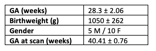
Fifteen preterm newborns were recruited at Geneva University Hospital (Figure 1). Informed consent was obtained from the parents prior to participation in the study. All subjects underwent musical intervention five times per week (mean number: 25 ± 8.9) during hospitalization. Finally, at term-equivalent age (TEA), we performed the MRI scan.
Image Acquisition
All infants were fed and MR-compatible headphones were used to protect them from noise. We acquired two runs (run 1, run 2) of 8 minutes of rs-fMRI data (300 volumes) (EPI, TR=1600 ms, TE=30 ms) on a Siemens 3T scanner. Between these two rs-fMRI runs, a task-based fMRI run was performed where the preterm infants listened to the same music used for the musical intervention during the hospitalization. Furthermore, a T2*-weighted structural image was acquired (TR=4990 ms, TE=151 ms).
Preprocessing
The data were first realigned and then co-registered using SPM8. Next, all volumes with a frame-wise displacement (FD) greater than 0.5 mm or with a rate of BOLD signal changes across the entire brain (DVARS) greater than 3% were removed, along with the previous and the two subsequent images. The remaining images were included in the analysis (1st run: mean: 270, 2nd run: 260).
Network Construction
To extract regional time-series from the rs-fMRI data, the UNC neonatal atlas7 is first registered to each subject’s space using Advanced Normalization Tools8. Next, the average signal of each ROI is calculated considering only grey matter voxels. The connectome of each subject is constructed using Accordance9 resulting in one connectome per run for each subject. The upper triangular of the connectivity matrix is vectorized resulting in a 4005-dimensional vector and finally, connections that are zero for more than 8 subjects are removed.
Statistical Analysis
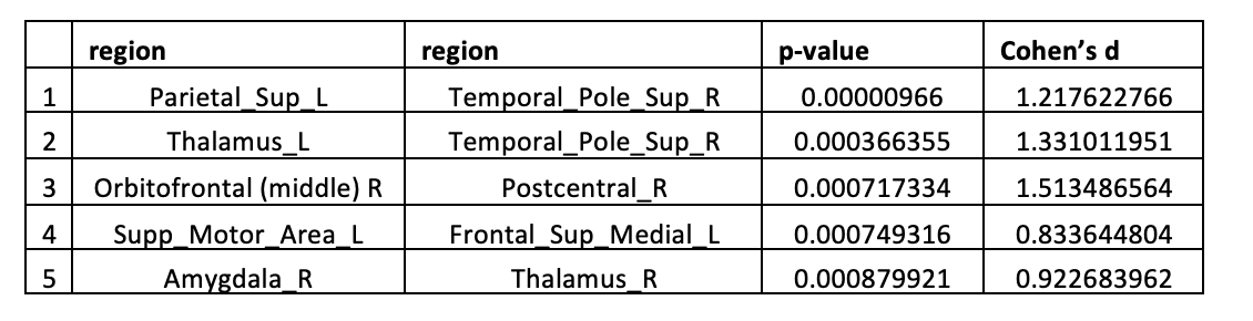
To unveil changes in functional connectivity evoked by the familiar musical stimuli presented between the rs-fMRI runs, paired t-tests were performed on each connection. Cohen’s d metric was computed to measure the effect size since the p-value significance threshold was set to 0.001 (uncorrected) due to the insufficient statistical power (n=15).
Results
Connection-wise Analysis:Paired t-tests results
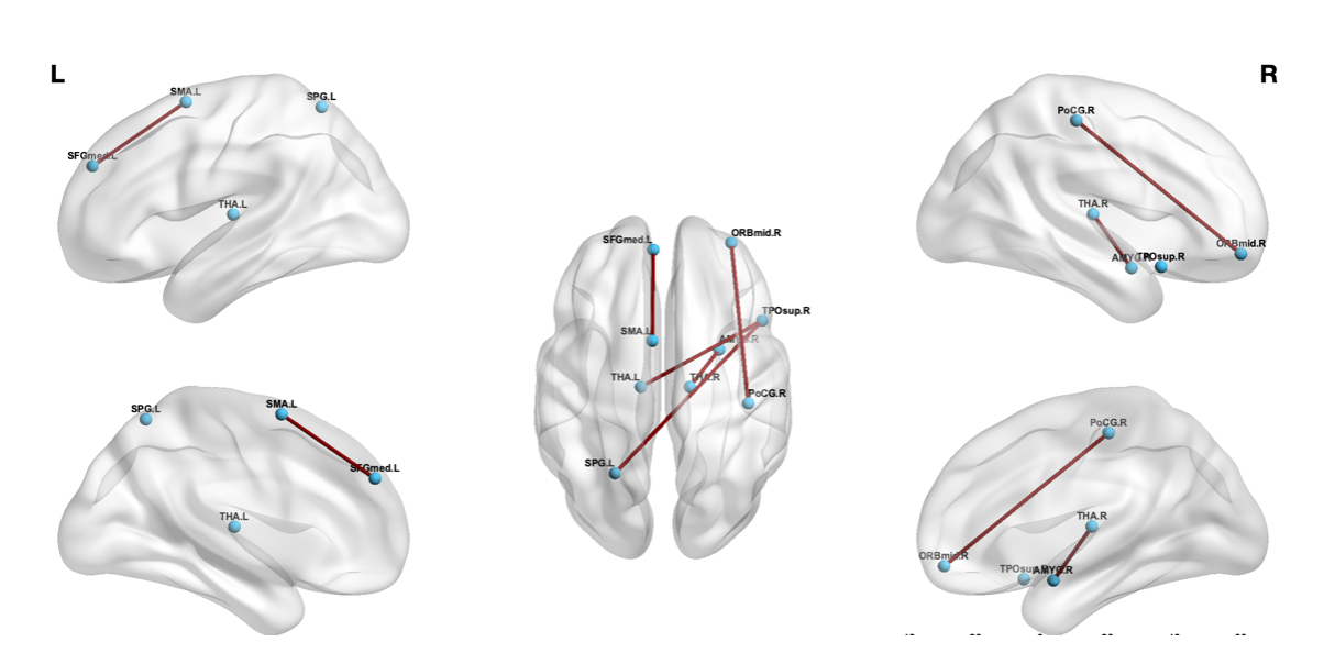
In this section, we report the connections that were stronger in run 2 compared to run 1 (Figure 2 and 3).
Discussion & Conclusion
The aim of this study was to explore functional connectivity changes in preterm newborns evoked by a familiar musical stimuluspreviously heard during the intervention in NICU. We hypothesized that after the presentation of the familiar musical stimulus, the functional connectivity in key areas involved in music processing might be increased.
Our results show that listening again to a known music increases functional connectivity between brain regions implicated in music processing, including the Temporal pole, Thalamus, and Sensorimotor areas; as well as brain regions implicated in music-evoked emotions such as Amygdala and Orbitofrontal cortex.10,11
Previously, using a Psychophysiological Interaction Analysis, we observed an increased functional connectivity between regions implicated in familiar and pleasant music processing, when listening again to the music heard during early music intervention.4 Here, we further show that increased functional connectivity linked to this familiar and pleasant music listening is still present at rest few minutes after.
Thus, listening to a familiar and pleasant music increases resting state functional connectivity between brain regions implicated in music and emotion processing and this modulation of resting state functional connectivity can be observed few minutes after music listening.
Acknowledgements
This study was supported by the Swiss National Science Foundation n°32473B_135817/1 and the foundation Prim'enfance. The authors thank all nurses implicated as well as all the parents and babies. We also thank Division of ENT, the Plateforme de Recherche de Pédiatrie and the Centre for Biomedical Imaging (CIBM) of the University Hospital of Geneva for their support.References
1. M. Delobel-Ayoub, C. Arnaud, M. White-Koning, C. Casper, V. Pierrat, M. Garel, A. Burguet, J. Roze, J. Matis, J. Picaud, M. Kaminski, and B. Larroque. Behavioral problems and cognitive performance at 5 years of age after very preterm birth: The EPIPAGE study. Pediatrics, 123(6), 2009.
2. C. Arpino, E. Compagnone, M. Montanaro, D. Cacciatore, A. De Luca, A. Cerulli, S. Di Girolamo, and P. Curatolo. Preterm birth and neurodevelopmental outcome: a review. Childs. Nerv. Syst., 26(9), 2010.
3. L. Doyle and P. Anderson. Adult outcome of extremely preterm infants. Pediatrics, 126(2), 2010.
4. L. Lordier, S. Loukas, F. Grouiller, A. Vollenweider, L. Vasung, D. Meskaldij, F. Lejeune, M. Pittet, C. Borradori-Tolsa, F. Lazeyras, D. Grandjean, D. Van De Ville, and P. Huppi. Music processing in preterm and full-term newborns: A psychophysiological interaction (PPI) approach in neonatal fMRI. NeuroImage, 2018.
5. O. Sporns, T. Giulio, and K. Rolf. The Human Connectome: A Structural Description of the Human Brain. PLoS Comput Biol, 1(4), 2005.
6. A. Fornito, A. Zalesky, and M. Breakspear. The connectomics of brain disorders. Nat Rev Neurosci., 16(3), 2015.
7. F. Shi, P. T. Yap, G. Wu, H. Jia, J. H. Gilmore, W. Lin, D. Shen, "Infant Brain Atlases from Neonates to 1- and 2 year-olds", PLOS ONE, 6(4), 2011.
8. B. B. Avants, N. J. Tustison, G. Song, P. A. Cook, A. Klein, J. C. Gee, A reproducible evaluation of ANTs similarity metric performance in brain image registration, NeuroImage, 54(3), 2011.
9. D. Meskaldji, S. Morgenthaler and D. Van De Ville, New measures of brain functional connectivity by temporal analysis of extreme events, 2015 IEEE 12th International Symposium on Biomedical Imaging (ISBI), 2015.
10. T. Särkämö, P. Ripollés, H. Vepsäläinen, et al. Structural changes induced by daily music listening in the recovering brain after middle cerebral artery stroke: a voxel-based morphometry study. Front Hum Neurosci. 2014.
11. S. Koelsch, Brain correlates of music-evoked emotion. Nature Reviews Neuroscience, 2014.
Figures