0043
Anomalous relationship between sensorimotor GABA levels and cortical excitability in children with Attention-deficit/hyperactivity disorder1Radiology, University of Calgary, Calgary, AB, Canada, 2Child and Adolescent Imaging Research Program, Alberta Children's Hospital Research Institute, University of Calgary, Calgary, AB, Canada, 3Hotchkiss Brain Institute, University of Calgary, Calgary, AB, Canada, 4Division of Pediatric Neurology, Cincinnati Children's Hospital Medical Center and University of Cincinnati, Cincinnati, OH, United States, 5Department of Radiology, Cincinnati Children's Hospital Medical Center and University of Cincinnati, Cincinnati, OH, United States, 6Russell H. Morgan Department of Radiology and Radiological Science, Johns Hopkins School of Medicine, Baltimore, MD, United States, 7F.M. Kirby Research Center for Functional Brain Imaging, Kennedy Krieger Institute, Baltimore, MD, United States, 8Center forNeurocognitive and Imaging Research, Kennedy Krieger Institute, Baltimore, MD, United States, 9Department of Neurology, Johns Hopkins School of Medicine, Baltimore, MD, United States, 10Department of Behavioral Science and Psychiatry, Johns Hopkins School of Medicine, Baltimore, MD, United States
Synopsis
Reduced inhibition as shown though GABA-edited MRS and TMS measures have been suggested to underlie some of the symptomology of ADHD. In this study we apply GABA-MRS and single- and paired-pulse TMS in ADHD and typically developing cohorts to better understand altered inhibition in ADHD. We show that SICI approaches 1 with increasing GABA across both groups. Secondly, we show a convergence in SICI and single-pulse TMS responses with increasing GABA. Last, we show different factors modulate the TMS responses between ADHD and control suggesting there are additional differences in GABAergic inhibition between these two groups.
Introduction
Attention-deficit/hyperactivity disorder (ADHD) is a neurodevelopmental disorder defined by inattention and impulsive, hypermotoric behaviors. Previous work has shown reduced inhibition in ADHD as indexed by (a) lower levels of the inhibitory neurotransmitter GABA, measured by GABA-edited MRS [1] and (b) reduced Transcranial Magnetic Stimulation (TMS)-evoked Short Interval Cortical Inhibition (SICI) [2].
TMS can be used to interrogate cortical excitation and inhibition. Single supra-threshold TMS pulses over motor cortex (M1) yield motor evoked potentials (MEPs) whose amplitudes reflect cortical excitability. Short interval (3 msec) paired sub- and supra-threshold pulses yield smaller amplitude MEPs, i.e. SICI, mediated by GABA-A receptors [3].
This study examines the relationship between MRS-measured GABA and TMS evoked responses in children with ADHD and typically developing children (TDC). The objective was to determine the neurobiology of altered inhibition in children with ADHD. Unexpected findings allow for a novel description of GABAergic inhibition in ADHD and typical development.
Methods
Participants: Across two sites, 131 children ages 8 to 12 years (were recruited: 66 ADHD, 65 TDC; 65% male; 62% white, 21% black, 7% Asian, 11% biracial; 10% Hispanic).
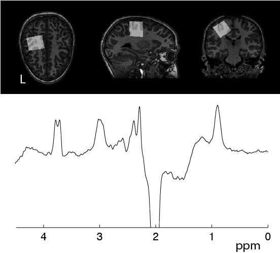
MRS: MR scanning was performed at 3T (Philips) and included T1-weighted imaging (MPRage, TR/TE=8 ms/3.7 ms, 1 mm3 isotropic voxels) for voxel placement and segmentation. GABA data were collected in the left sensorimotor cortex (left SM1) in 3×3×3 cm3 voxels using MEGA-PRESS (TR/TE=2s/68 ms, 14 ms editing pulses at 1.9 ppm and 7.46 ppm, 320 averages, 16-step phase cycle, and 8 unsuppressed water scans) and was quantified using Gannet [4,5] (Figure 1).
TMS: TMS (Magstim Bistim 200) was applied to left primary motor cortex using a round 90mm coil placed optimally near the vertex. MEPs were measured with electromyography in the right first dorsal interosseous muscle (Signal v6; CED 1401; Cambridge UK). MEP resting motor threshold (RMT) was determined using standard procedures.[6] Twenty single pulses (applied at 1.2*RMT) and paired pulses (first applied at 0.6*RMT, 3 msec interstimulus interval, second pulse applied at 1.2*RMT) were administered in randomized order, with SICI calculated as the mean MEP from the paired pulse over mean single pulse MEPs (standard method of means).
Analysis: Bivariate correlations were evaluated between SICI (method of means) and
GABA. Separately, relationships of single and paired MEP amplitudes and GABA
were modeled using repeated-measures generalized linear mixed models, a highly novel
approach, taking into account variability across TMS trials. Models accounted
for age, sex, site, pulse-type, diagnosis, and subtle artifact quantified using
the 200 msec prior to TMS, which is assumed to be noise.
Results
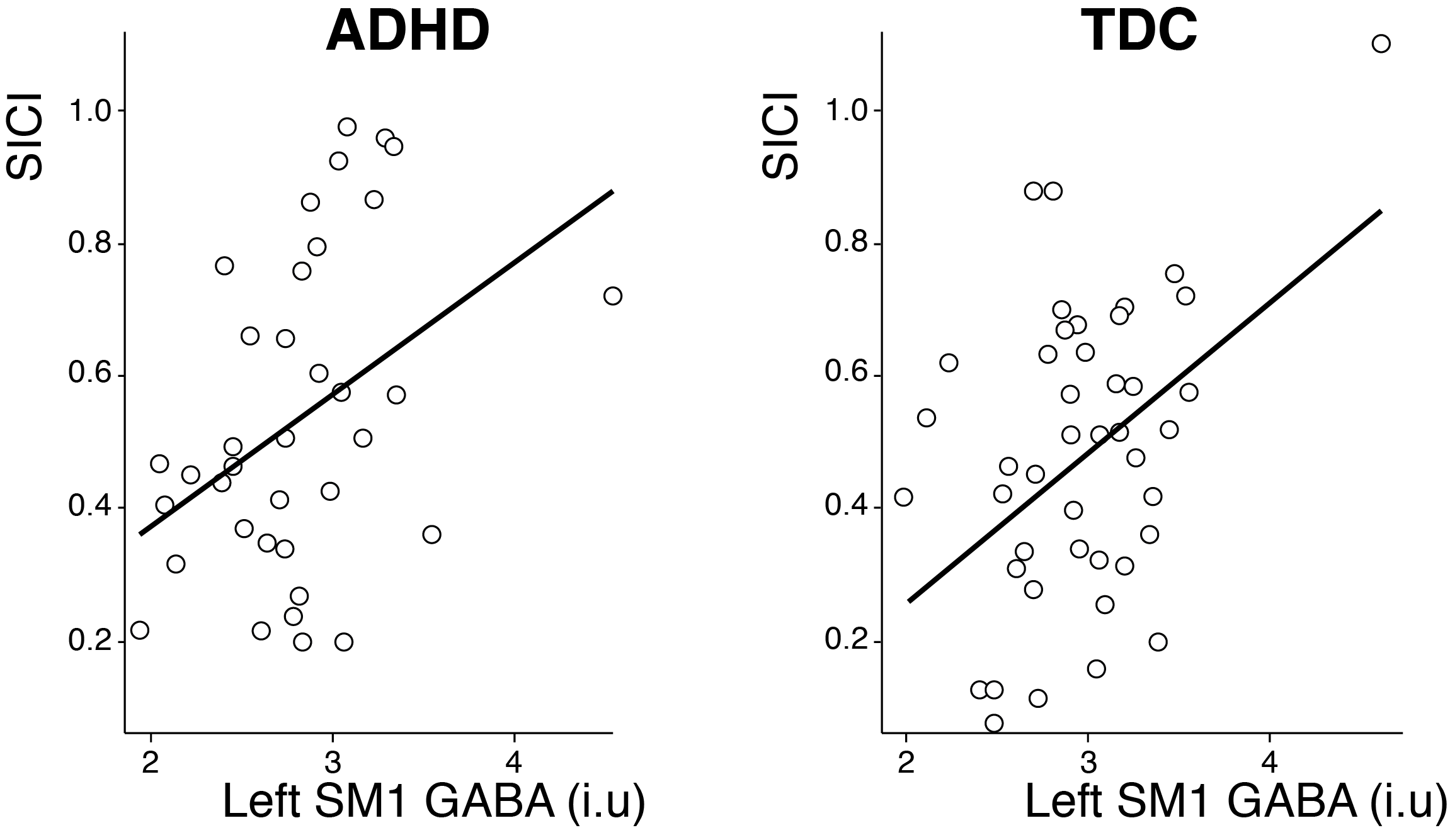
Left SM1 GABA did not differ between ASD and TDC groups (p = 0.21). There was significantly less SICI in ADHD (43%, 95% CI 37-49%) compared to TDC (53%, 95% CI 47-59%) (p = 0.028). In both groups, higher SM1 GABA levels correlated significantly with larger SICI ratios (less inhibition) (Figure 2).
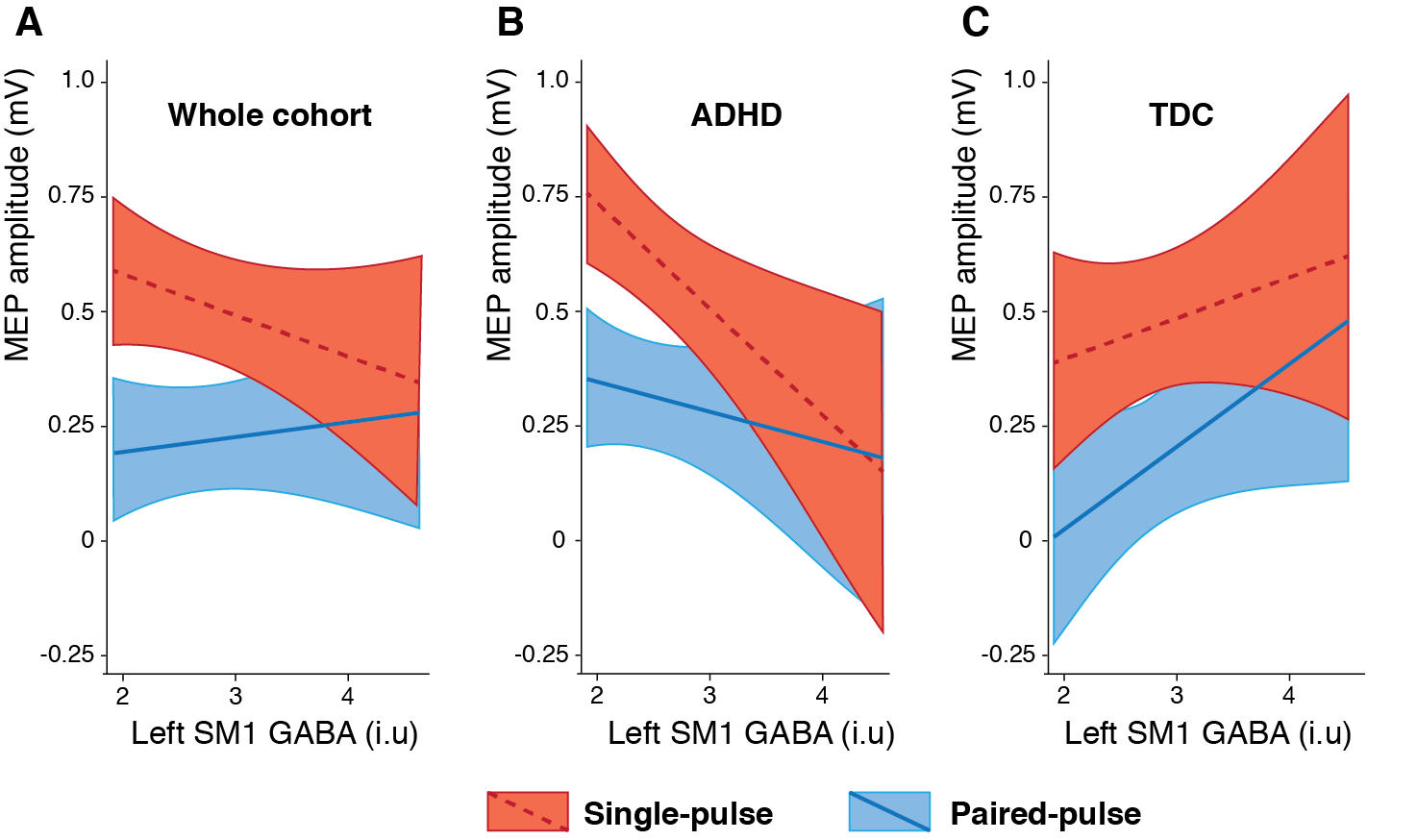
Mixed models demonstrated a highly significant interaction between pulse type and SM1 GABA (t1502 = 4.1, p < 0.0001), as paired and single pulse MEP amplitudes diverge with lower and converge with higher SM1 GABA levels (Figure 3A). This interaction remained significant after stratifying by diagnosis; however the MEP/GABA slopes are inverted (ADHD p = 0.0003, negative slope; TDC p = 0.02, positive slope) (Figure 3B and 3C).
Discussion
Simple correlational analysis showed that children with lower levels of GABA (measured in left sensorimotor cortex with GABA-edited MRS) had more physiological inhibition (measured in left motor cortex with TMS-evoked SICI). To our knowledge this is the first time this result has been reported.
Evaluation of SICI components – inhibitory paired versus single pulse MEP amplitudes – revealed that among children with higher GABA levels, paired pulse amplitudes are larger relative to single pulse amplitudes. These data show a convergence; among children with higher GABA levels, the difference between single and paired-pulse MEP size narrows (Figure 3a). However, the overall relationships between MEP amplitudes and GABA levels differs by diagnosis, i.e. shown in Figures 3B and 3C the slopes of the MEP-GABA plots are positive in TDC but negative in ADHD. This suggests an anomalous relationship in children with ADHD between cortical physiology and available GABA influenced by development and cortical metabolism, and merits further investigation.
Acknowledgements
This project was supported by funding from R01 MH078160 and R00MH107719.References
[1] Edden, Crocetti, Zhu, Gilbert Mostofsky. Reduced GABA Concentration in Attention-Deficit/Hyperactivity Disorder. Arch Gen Psychiatry. 2012; 69: 750 – 753.
[2] Gilbert, Isaacs, Augusta, Macneil, Mostofsky. Motor cortex inhibition: a marker of ADHD behaviour and motor development in children. Neurology. 2011; 76: 615-621.
[3] Stagg, Bestmann, Constantinescu, Moreno, Allman, Mekle, Woolrich, Near, Johansen-Berg, Rothwell. Relationship between physiological measures of excitability and levels of glutamate and GABA in the human motor cortex. J Physiol. 2011; 589:.23: 5854-5855.
[4] Edden, Puts, Harris, Barker, Evans. Gannet: A batch-processing tool for the quantitative analysis of gamma-aminobutyric acid-edited MR spectroscopy spectra. J Magn Reson Imaging. 2014; 40: 1445-1452.
[5] Harris, Puts, Edden. Tissue correction for GABA-edited MRS: Considerations of voxel composition, tissue segmentation and tissue relaxations. J Magn Reson Imaging. 2015; 42: 1431-1410.
[6] Mills, Nithi. Corticomotor threshold to magnetic stimulation: normal values and repeatability. Muscle Nerve 1997;20:570 –576.
Figures