0035
Changes in functional brain connectivity associated with the development of executive functions during early childhood1Warren Alpert Medical School, Department of Pediatrics, Brown University, Providence, RI, United States, 2Women and Infants Hospital of Rhode Island, Providence, RI, United States, 3MNCHD&T, Bill & Melinda Gates Foundation, Seattle, WA, United States
Synopsis
Task-based functional MRI (fMRI) has been commonly used to study executive functions but may be difficult to implement for children younger than 5-year-old. In this work, resting sate fMRI is combined with in-house tablet-based tasks which evaluate reaction and inhibition control in children from 2 to 5 years of age. The aim is to determine differences in brain connectivity related to the children’s performance of these two tasks. Preliminary results demonstrated the importance of the language, visual and attention networks during the reaction task and asymmetrical changes in frontal parietal and sensorimotor network connectivity associated with the development of inhibition control.
Introduction
Executive function (EF) is the ability to control and regulate actions and thoughts1. It includes processes such as working memory, self-regulation, and inhibitory control. To date, most evaluations of EF rely on parents’ reports such as the Behavior Rating Inventory of Executive Function Preschool (BRIEF-P) form, which may not capture the development of executive skills to its full expression. Recently we developed a tablet-based assessment consisting of a reaction time task and an inhibition task. McLean et al.2 found strong correlations of the children’s performance during reaction and inhibition with age but not with other standard cognitive measures (BRIEF-P, WISC). These results underline the ability of our tablet-based task to assess rapid changes in EF during early childhood that is not captured by traditional assessments.
EF has been previously studied using task-based functional MRI (fMRI) scans3, which can be difficult to adapt for testing in young children. Instead, multiple studies have relied on resting state functional MRI4–6. Among them, Reineberg et al.6 investigated differences in brain connectivity in relation to individual performances during different EF behavioral tasks. However, to our best knowledge, resting state fMRI has not been applied to characterize EF-related brain connectivity differences in children, especially at a young age (2-5 year old).
Methods
Resting state functional data and structural scans were acquired on 24 typically developing children (range=21-70 mo; mean=47.3 mo; 9 females) during natural sleep on a 3T Trio scanner in accordance with the local IRB. Resting state fMRI data were acquired with the following parameters: TE=34 ms, TR=2.5 s, voxel resolution: 3x3x6 mm3, 32 slices. FMRI data were first preprocessed (realignment, motion correction, scrubbing) with the MATLAB CONN toolbox7 and registered to our child study template using FSL FLIRT8 and ANTS9. In addition, the children were assessed using the tablet-based tasks in which a reaction and an inhibition task were implemented. In the reaction task, the child was asked to click quickly on the image of a dog appearing on the screen. The inhibition task was similar, but images of a ball were introduced into the sequence. The child was asked to click when a dog appeared, but not on the ball. The measures of interest were the reaction time from the reaction task and the number of balls clicked (errors) from the inhibition task. Group level ROI-to-ROI connectivity analysis was performed with CONN to determine differences in brain connectivity with the two EF measures. Gender and age were defined as covariates in the analysis. 32 ROIs corresponding to commonly reported networks were used in the analysis. Significant differences in network connectivity were defined for p ≤.05 FDR seed-level corrected for multiple comparisons.Results
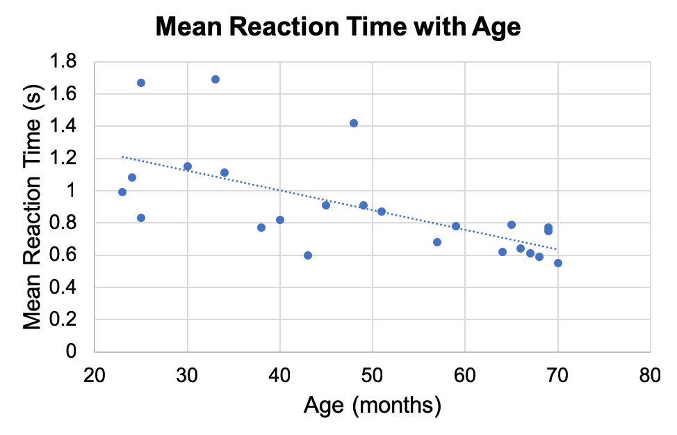
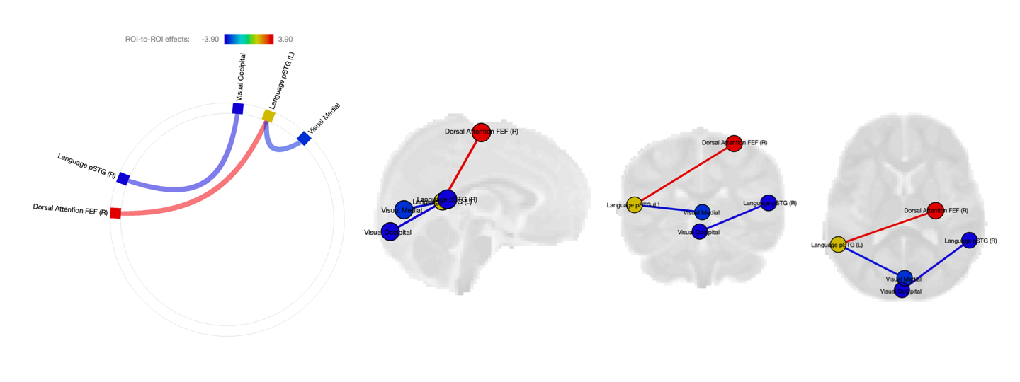
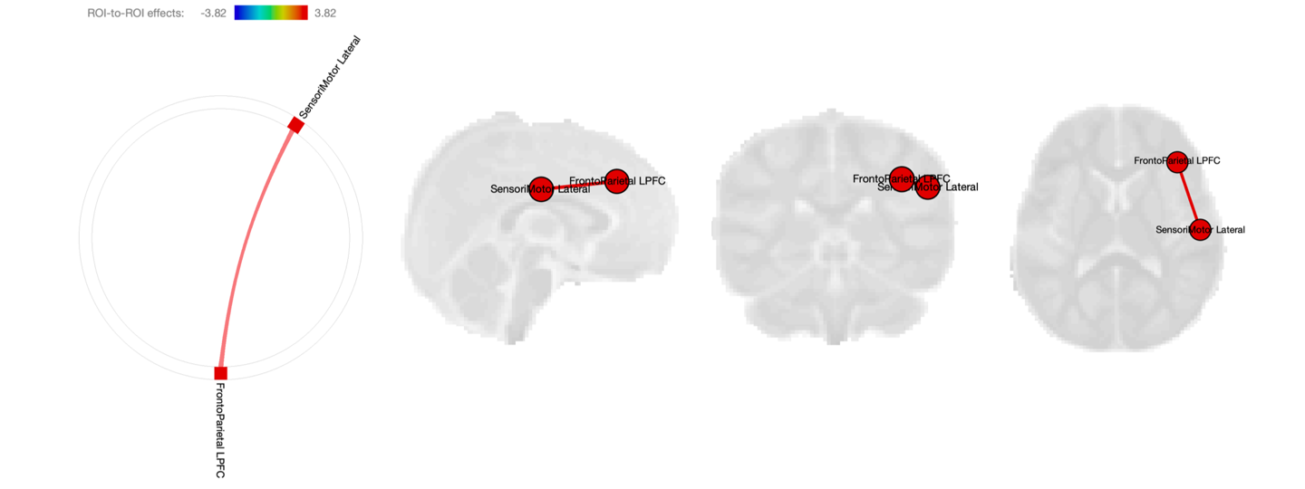
Children are faster with age as EF develops (Figure 1). Figure 2 suggests that slower children (higher reaction time) have reduced connectivity between the visual and language networks but stronger connectivity between the dorsal attention network and language network. An increased connection between the right lateral sensorimotor and the right frontal parietal network is observed with a higher number of balls clicked (errors) during the inhibition task (Figure 3). No sex differences in network connectivity were observed in our dataset.Discussion
The differences in brain connectivity suggest the importance of language, visual and attention networks during the reaction task. The involvement of the language network alongside the development of EF has been reported in previous studies10,11, where children with language impairments also showed persistent EF deficits. Furthermore, stronger connectivity was observed between the visual and language networks in children who were quicker during the task. Differences in the frontoparietal network connectivity are correlated to the inhibition task performance. This finding is in agreement with previous work demonstrating the role of the prefrontal cortex in inhibitory control12–14. The observed asymmetry could suggests right hemisphere dominance15 at a young age. Of note is also the stronger positive connectivity between the frontoparietal network and sensorimotor areas associated with poorer inhibition control. These results could indicate frontoparietal and sensorimotor networks changing with the development of inhibition control during the first 5 years of life.Conclusion
Better understanding how brain connectivity changes with the development of executive functions can help characterize healthy child development as well as aid early EF delay detection. The developed tablet-based task combined with resting state fMRI provides a suitable approach to study EF in younger children. Our preliminary results suggest that the language, visual, dorsal attention network and the frontoparietal and sensorimotor network play an important role at a young age in the development of EF, evaluated by the reaction and inhibition tasks that are presented in this work.Acknowledgements
No acknowledgement found.References
1. Diamond, A. Executive Functions. Annu. Rev. Psychol. 64, 135–168 (2013). 2. Mclean, R. et al. Development of novel tablet-based measures of early learning and developing cognitive skills in early childhood: Preliminary feasibility and validity. in Proceedings of Society for Neuroscience Meeting, San Diego, CA, Nov 3-7 2018 (2018). 3. McKenna, R., Rushe, T. & Woodcock, K. A. Informing the Structure of Executive Function in Children: A Meta-Analysis of Functional Neuroimaging Data. Front. Hum. Neurosci. 106, 124-134, (2017). 4. Reineberg, A. E., Andrews-Hanna, J. R., Depue, B., Friedman, N. P. & Banich, M. T. Resting-state Networks Predict Individual Differences in Common and Specific Aspects of Executive Function. NeuroImage 104, 69–78 (2015). 5. Kornfeld, S. et al. Resting-state connectivity and executive functions after pediatric arterial ischemic stroke. NeuroImage Clin. 17, 359–367 (2017). 6. Reineberg, A. E., Gustavson, D. E., Benca, C., Banich, M. T. & Friedman, N. P. The Relationship Between Resting State Network Connectivity and Individual Differences in Executive Functions. Front. Psychol. 9, 1600 (2018). 7. Whitfield-Gabrieli, S. & Nieto-Castanon, A. Conn: a functional connectivity toolbox for correlated and anticorrelated brain networks. Brain Connect. 2, 125–141 (2012). 8. Smith, S. M. et al. Advances in functional and structural MR image analysis and implementation as FSL. NeuroImage 23 Suppl 1, S208-219 (2004). 9. Avants, B. B. et al. The Insight ToolKit image registration framework. Front. Neuroinformatics 8, 44 (2014). 10. Gooch, D., Thompson, P., Nash, H. M., Snowling, M. J. & Hulme, C. The development of executive function and language skills in the early school years. J. Child Psychol. Psychiatry 57, 180–187 (2016). 11. Yang, H.-C. & Gray, S. Executive Function in Preschoolers with Primary Language Impairment. J. Speech Lang. Hear. Res. 60, 379–392 (2017). 12. Best, J. R. & Miller, P. H. A Developmental Perspective on Executive Function. Child Dev. 81, 1641–1660 (2010). 13. Houdé, O. & Borst, G. Measuring inhibitory control in children and adults: brain imaging and mental chronometry. Front. Psychol. 5, 616 (2014). 14. Moriguchi, Y. Neural Mechanisms of Executive Function Development during Early Childhood. Executive Function (2017). 15. Garavan, H., Ross, T. J. & Stein, E. A. Right hemispheric dominance of inhibitory control: An event-related functional MRI study. Proc. Natl. Acad. Sci. U. S. A. 96, 8301–8306 (1999).Figures