0032
Music in preterm infants enhances maturation of neural pathways involved in emotion processing1Division of Development and Growth, Department of Pediatrics, University of Geneva, Geneva, Switzerland, 2Division of Development and Growth, Department of Pediatrics, Geneva University Hospitals, Geneva, Switzerland, 3Psychology, Yale University, New Haven, CT, United States, 4Center of BioMedical Imaging (CIBM), Ecole Polytechnique Fédérale de Lausanne (EPFL), Lausanne, Switzerland, 5Wellcome Centre for Integrative Neuroimaging (WIN) - Oxford Centre for Functional Magnetic Resonance Imaging of the Brain (FMRIB), University of Oxford, Oxford, United Kingdom, 6Sir Peter Mansfield Imaging Centre, School of Medicine, University of Nottingham, Nottingham, United Kingdom, 7Center of BioMedical Imaging (CIBM), University of Geneva, Geneva, Switzerland
Synopsis
Prematurity disrupts brain maturation during a critical period of development and music potentially enhances cognitive-socio-emotional pathways affected by prematurity. Using multi-modal MRI, we evaluated the structural impact of a music intervention during NICU stay in preterm infants’ brains, namely in WM through DTI ROI and tractography analysis and in amygdala through volumetric analysis. Overall, WM microstructural maturity was decreased in preterm control vs full-term newborns. In comparison to preterm control, preterm exposed to music demonstrate improved WM maturation in uncinate fasciculus, external capsule/claustrum/extreme capsule and larger amygdala volumes, proving a structural effect of music intervention on emotional processing neural pathways.
INTRODUCTION
Prematurity disrupts brain maturation by exposing the developing brain to different noxious stimuli present in the neonatal intensive care unit (NICU) and depriving it from meaningful sensory inputs,1,2 during a critical period of brain development.3 Evidence has proven that preterm birth is accompanied by structural brain alterations, including cortical and subcortical volumetric changes and white matter (WM) abnormalities, correlating with later neurodevelopmental impairments affecting up to 40% of very preterm infants (born <32 weeks gestational age).4-11 Musicotherapy has been used in NICU as an approach for meaningful sensory stimulation, relevant for activity-dependent brain plasticity. In fact, music listening triggers neural substrates implied in cognitive-socio-emotional processing12-15 and might thus influence networks formed early in development and affected by prematurity. Using multi-modal Magnetic Resonance Imaging (MRI), we aimed to study the impact of a music intervention on premature infants’ structural brain development.METHODS
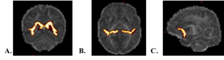
15 full-term and 30 very preterm newborns at term-equivalent-age (15 without music exposure (PTC) and 15 exposed daily to music (PTM) during NICU stay) underwent an MRI (3T Siemens Prisma) exam comprising a Diffusion Tensor Imaging (DTI) (TE=84ms, TR= 7400ms, voxel size 2x2x2mm3, gradient applied in 30 non-collinear directions with b-value 1000 s/mm2 and one non-diffusion weighted image) and a T2-weighted image acquisition (TE=151ms, TR= 4990ms, voxel size 0.4x0.4x1.2mm3). DTI data were preprocessed using FSL v5.0.10 diffusion toolbox16,17 and distortions caused by motion and eddy currents were corrected using EDDY adapted for neonatal data.18,19 DTI scalar maps, including Fractional anisotropy (FA) and Mean Diffusivity (MD), were derived in order to evaluate WM microstructure, using two approaches: region-of-interest (ROI) and seed-based tractography analysis. Regarding ROI analysis, 20 ROI were manually drawn in a study template, generated with DTI-TK20 by means of normalization of all subject diffusion images, and back transformed to each subject space (figure 1). Mean FA and MD were computed for each region and also averaged across all 20 ROI per subject and compared between groups, using one-way ANCOVA correcting for gestational age at MRI (GA-MRI) with Bonferroni correction. FSL seed-based probabilistic tractography was performed for the following tracts: acoustic radiations, interhemispheric temporal callosal fibers and uncinate fasciculus (figure 2). Masks were defined for each tract in the subject diffusion space, where the probabilistic tractography was performed, according to published literature and atlas.21-23 Mean FA and MD were calculated per subject tract and compared between groups, using one-way ANCOVA (Bonferroni corrected) correcting for GA-MRI. Additionally, amygdala segmentation was manually performed by a single operator on the T2-weighted images using ITK-snap, and corrected by an expert neurosurgeon (figure 3). Segmentation was based on anatomical guidelines according to published literature and neonatal atlas.10,24 Amygdala volumes were compared between groups using one-way ANCOVA (Bonferroni corrected) correcting for intracranial cavity volume, GA-MRI and sex.RESULTS AND DISCUSSION
Considering
the average of all ROIs per subject, mean FA was significantly lower (p=0.0001)
and MD significantly higher (p=0.008) in PTC vs full-term newborns, showing
that preterm when at term-equivalent age present a decreased brain maturation
in comparison to full-term newborns; PTM values were not significantly
different from full-term newborns. When analyzing per region, PTM showed a
significantly higher FA vs PTC (p=0.01) in the ROI “ec” (external
capsule/claustrum/extreme capsule), where association fibers connecting regions
involved in music processing are located.
Tractography analysis of acoustic
radiations, which relay the auditory information from the thalamus to primary
auditory cortex, revealed that PTM had a higher mean FA than PTC (p=0.034), but
this group difference did not survive Bonferroni correction (p=0.068).
Regarding the interhemispheric temporal callosal fibers, which transmit the
auditory information between the hemispheres, PTC presented a significantly
lower FA (p=0.006) and higher MD (p=0.0001) in comparison to full-term group,
thus showing a decreased maturation vs full-term, whereas PTM music presented a
mean FA not significantly different from full-term newborns. Analysis of the
uncinate fasciculus, known to be involved in emotion regulation processes and
in music processing, evidenced a significantly higher FA in PTM vs PTC
(p=0.048), highlighting an effect of music intervention in the maturation of
this tract in preterm infants.
Lastly, the amygdala volumetric analysis showed
that PTC have a lower amygdala volume vs full-term infants (p=0.005), whereas
PTM had a significantly larger amygdala volume vs PTC (p=0.014), evidencing a
structural beneficial effect of the music intervention on amygdala volume in
preterm infants.
CONCLUSION
Overall, microstructural maturity
was decreased in preterm at term vs full-term newborns. Preterm infants exposed to music showed an
increased WM microstructural maturation in uncinate fasciculus, external
capsule/claustrum/extreme capsule and a larger amygdala volume, proving a
structural effect of music intervention on emotional processing pathways.
Acknowledgements
We acknowledge the Pediatrics Clinic Research Platform of HUG for their help and support, as well as all parents and their infants for their participation in this study.
Special acknowledgment to Dr. Samuel Sommaruga, neurosurgeon, for his contribution to amygdala segmentation correction.
This work was supported by the Center for Biomedical Imaging (CIBM) of the University and Hospitals of Geneva.
Research funded by Swiss National Science Foundation Program (324730-163084).
References
1. Radley JJ, Morrison JH. Repeated stress and structural plasticity in the brain. Ageing Res Rev 2005; 4(2): 271-87.
2. Kiss JZ, Vasung L, Petrenko V. Process of cortical network formation and impact of early brain damage. Curr Opin Neurol 2014; 27(2): 133-41.
3. Blencowe H, Cousens S, Chou D, et al. Born too soon: the global epidemiology of 15 million preterm births. Reproductive health 2013; 10 Suppl 1: S2-S.
4. Huppi PS, Maier SE, Peled S, et al. Microstructural development of human newborn cerebral white matter assessed in vivo by diffusion tensor magnetic resonance imaging. Pediatric research 1998; 44(4): 584-90.
5. Inder TE, Warfield SK, Wang H, Huppi PS, Volpe JJ. Abnormal cerebral structure is present at term in premature infants. Pediatrics 2005; 115(2): 286-94.
6. Thompson DK, Warfield SK, Carlin JB, et al. Perinatal risk factors altering regional brain structure in the preterm infant. Brain 2007; 130(Pt 3): 667-77.
7. Thompson DK, Adamson C, Roberts G, et al. Hippocampal shape variations at term equivalent age in very preterm infants compared with term controls: perinatal predictors and functional significance at age 7. Neuroimage 2013; 70: 278-87.
8. Anjari M, Srinivasan L, Allsop JM, et al. Diffusion tensor imaging with tract-based spatial statistics reveals local white matter abnormalities in preterm infants. Neuroimage 2007; 35(3): 1021-7.
9. Ball G, Boardman JP, Rueckert D, et al. The effect of preterm birth on thalamic and cortical development. Cereb Cortex 2012; 22(5): 1016-24.
10. Cismaru AL, Gui L, Vasung L, et al. Altered Amygdala Development and Fear Processing in Prematurely Born Infants. Front Neuroanat 2016; 10: 55.
11. Montagna A, Nosarti C. Socio-Emotional Development Following Very Preterm Birth: Pathways to Psychopathology. Frontiers in Psychology 2016; 7.
12. Koelsch S, Kasper E, Sammler D, Schulze K, Gunter T, Friederici AD. Music, language and meaning: brain signatures of semantic processing. Nature neuroscience 2004; 7(3): 302-7.
13. Popescu M, Otsuka A, Ioannides AA. Dynamics of brain activity in motor and frontal cortical areas during music listening: a magnetoencephalographic study. NeuroImage 2004; 21(4): 1622-38.
14. Koelsch S. Towards a neural basis of music-evoked emotions. Trends in cognitive sciences 2010; 14(3): 131-7.
15. Zatorre RJ, Peretz I, Penhune V. Neuroscience and Music ("Neuromusic") III: disorders and plasticity. Preface. Annals of the New York Academy of Sciences 2009; 1169: 1-2.
16. Smith SM, Jenkinson M, Woolrich MW, et al. Advances in functional and structural MR image analysis and implementation as FSL. Neuroimage 2004; 23: S208-S19.
17. Behrens TEJ, Woolrich MW, Jenkinson M, et al. Characterization and propagation of uncertainty in diffusion-weighted MR imaging. Magn Reson Med 2003; 50(5): 1077-88.
18. Andersson JLR, Sotiropoulos SN. An integrated approach to correction for off-resonance effects and subject movement in diffusion MR imaging. Neuroimage 2016; 125: 1063-78.
19. Bastiani M, Andersson JLR, Cordero-Grande L, et al. Automated processing pipeline for neonatal diffusion MRI in the developing Human Connectome Project. Neuroimage 2018.
20. Zhang H, Yushkevich PA, Alexander DC, Gee JC. Deformable registration of diffusion tensor MR images with explicit orientation optimization. Med Image Anal 2006; 10(5): 764-85.
21. Oishi K, Faria AV, Zijl PV, Mori S. MRI Atlas of Human White Matter 2nd ed: Academic Press 2010.
22. Adibpour P, Dubois J, Dehaene-Lambertz G. Right but not left hemispheric discrimination of faces in infancy. Nat Hum Behav 2018; 2(1): 67-79.
23. Akazawa K, Chang L, Yamakawa R, et al. Probabilistic maps of the white matter tracts with known associated functions on the neonatal brain atlas: Application to evaluate longitudinal developmental trajectories in term-born and preterm-born infants. Neuroimage 2016; 128: 167-79.
24. Gousias IS, Edwards AD, Rutherford MA, et al. Magnetic resonance imaging of the newborn brain: Manual segmentation of labelled atlases in term-born and preterm infants. Neuroimage 2012; 62(3): 1499-509.
Figures