0028
Hippocampal segmentation for brains with extensive atrophy using three-dimensional convolutional neural networksMaged Goubran1, Edward Ntiri1, Hassan Akhavein1, Melissa Holmes1, Sean Nestor1, Ramirez Joel1, Sabrina Adamo1, Fuqiang Gao1, Christopher Scott1, Anne Martel1, Walter Swardfager1, Mario Masellis1, Rick Swartz1, Bradley MacIntosh1, and Sandra Black1
1Sunnybrook Research Institute, Toronto, ON, Canada
Synopsis
Obtaining hippocampal volumes through manual segmentation requires an expert and is time consuming. Automated segmentation techniques would benefit from user-friendly and publicly accessible to tools, and robust results in the face of brain diseases. To accomplish these objectives, we trained a 3D convolutional neural network to segment the hippocampus automatically. Our algorithm was more accurate and time efficient compared to 4 publicly available state-of-the-art methods when considering a wide range of patient groups. Thus, we present a new method for obtaining hippocampal volumes, an important biomarker in aging, disease, and dementia.
INTRODUCTION
Hippocampal volumetry is a critical biomarker of neurodegeneration as this volume can predict cognitive decline and dementia risk 1- 6. However, segmenting the hippocampus is challenging due the complex and variable anatomy that is differentially affected in disease 7. Manual segmentation of the hippocampus is time consuming and may suffer in reproducibility across different raters. Automated methods of obtaining hippocampal volumetry are limited because 1) the algorithms are not publicly available, 2) they are unable to handle individuals with large brain atrophy, vascular disease, or lesions and strokes, and/or 3) require large computational time and computational knowledge 8-11.METHODS
We trained a 3D convolutional neural network using 259 bilateral manually delineated segmentations collected from 3 studies, which included multiple sites and different scanners (pulse sequence and field strength differences). Our test dataset consisted of difficult cases to segment due to varying levels of brain atrophy, vascular disease, lesions and strokes in this elderly population. Our algorithm, HyperMatter, was validated against four other techniques: HippoDeep, Freesurfer, SBHV and FIRST. We evaluated algorithm performances by comparison against manual segmentation using the following metrics: 1) the Pearson R. correlation coefficient of the volumes, 2) the Dice similarity coefficient, and 3) the Jaccard coefficient. Further validation of our model was performed to simulate clinical grade or challenging scans acquired with 1) decreases in resolution, 2) addition of noise, and 3) cropping of FOV.RESULTS
While all tested techniques provided significant correlations between predicted and manual volumes, our model generated the highest agreement with ground truth labels (r=0.95) and the lowest number of outliers (Figure 1, A). The distributions of Dice and Jaccard coefficients between ground truth manual labels of the hippocampus and predicted segmentations are presented in Figure 1, B. Our model outperformed the other state-of-the-art techniques by a margin bigger than 5%, generating an average Dice of 0.89 in these difficult test cases (Figure 2) . It was two orders of magnitude faster than some of the tested techniques, segmenting the hippocampi in an average of 14 seconds. The cases with the highest and lowest Dice coefficients from the test dataset between manual hippocampal segmentations and our prediction (0.92 and 0.80) are presented in Figure 3. HyperMatter proved to be robust to downsampling of images to 2x and cropping FOV in the Superior-Inferior plane by 30% but was more sensitive to the addition of a large amount (sigma) of speckle noise producing a Dice coefficient drop of around 14% (Figure 4).DISCUSSION
HyperMatter outperformed other available automated segmentations, including difficult, harder to segment cases due to large atrophy on average, smaller structure volumes, increased CSF, and presence of white matter lesions. We achieved high Dice scores of around 0.89 for the whole hippocampus, higher than any other previous automated segmented, and approaching expert manual raters 12. This might be because our hippocampal model was trained on multiple segmentation protocols which have slightly different border definitions and a lower inter-rater ICC than other manual segmentations. It would be optimal to test it on data with manual ground truth segmentations from other studies not part of the training set. Future work would include hippocampal subfield segmentation; however, this is currently limited due to availability of manual subfield segmentations on a large cohort and lack of histological validation.Acknowledgements
This study was funded by the Canadian Institute for Health Research (CIHR) MOP Grant #13129 and the L.C Campbell FoundationReferences
- Courchesne, E., Chisum, H.J., Townsend, J., Cowles, A., Covington, J., Egaas, B., Harwood, M., Hinds, S., Press, G.A., 2000. Normal brain development and aging: quantitative analysis at in vivo MR imaging in healthy volunteers. Radiology 216, 672–682.
- Jack, C.R., Jr, Petersen, R.C., Xu, Y., O’Brien, P.C., Smith, G.E., Ivnik, R.J., Tangalos, E.G., Kokmen, E., 1998. Rate of medial temporal lobe atrophy in typical aging and Alzheimer’s disease. Neurology 51, 993–999.
- Jack, C.R., Petersen, R.C., Xu, Y., O’Brien, P.C., Smith, G.E., Ivnik, R.J., Boeve, B.F., Tangalos, E.G., Kokmen, E., 2000. Rates of hippocampal atrophy correlate with change in clinical status in aging and AD. Neurology 55, 484–490.
- Rusinek, H., De Santi, S., Frid, D., Tsui, W.-H., Tarshish, C.Y., Convit, A., de Leon, M.J., 2003. Regional brain atrophy rate predicts future cognitive decline: 6-year longitudinal MR imaging study of normal aging. Radiology 229, 691–696.
- Scahill, R.I., Frost, C., Jenkins, R., Whitwell, J.L., Rossor, M.N., Fox, N.C., 2003. A longitudinal study of brain volume changes in normal aging using serial registered magnetic resonance imaging. Arch. Neurol. 60, 989–994.
- Sullivan, E.V., 2002. Differential Rates of Regional Brain Change in Callosal and Ventricular Size: a 4-Year Longitudinal MRI Study of Elderly Men. Cereb. Cortex 12, 438–445.
- Goubran, M., Rudko, D.A., Santyr, B., Gati, J., Szekeres, T., Peters, T.M., Khan, A.R., 2014. In vivo normative atlas of the hippocampal subfields using multi-echo susceptibility imaging at 7 Tesla. Hum. Brain Mapp. 35, 3588–3601.
- Fischl, B., Salat, D.H., Busa, E., Albert, M., Dieterich, M., Haselgrove, C., van der Kouwe, A., Killiany, R., Kennedy, D., Klaveness, S., Montillo, A., Makris, N., Rosen, B., Dale, A.M., 2002a. Whole brain segmentation: automated labeling of neuroanatomical structures in the human brain. Neuron 33, 341–355.
- Iglesias, J.E., Augustinack, J.C., Nguyen, K., Player, C.M., Player, A., Wright, M., Roy, N., Frosch, M.P., McKee, A.C., Wald, L.L., Fischl, B., Van Leemput, K., Alzheimer’s Disease Neuroimaging Initiative, 2015a. A computational atlas of the hippocampal formation using ex vivo, ultra-high resolution MRI: Application to adaptive segmentation of in vivo MRI. Neuroimage 115, 117–137.
- Nestor, S.M., Gibson, E., Gao, F.-Q., Kiss, A., Black, S.E., Alzheimer’s Disease Neuroimaging Initiative, 2013. A direct morphometric comparison of five labeling protocols for multi-atlas driven automatic segmentation of the hippocampus in Alzheimer’s disease. Neuroimage 66, 50–70.
- Thyreau, B., Sato, K., Fukuda, H., Taki, Y., 2018. Segmentation of the hippocampus by transferring algorithmic knowledge for large cohort processing. Med. Image Anal. 43, 214–228. 1
- Boccardi, M., Bocchetta, M., Morency, F.C., Collins, D.L., Nishikawa, M., Ganzola, R., Grothe, M.J., Wolf, D., Redolfi, A., Pievani, M., Antelmi, L., Fellgiebel, A., Matsuda, H., Teipel, S., Duchesne, S., Jack, C.R., Jr, Frisoni, G.B., EADC-ADNI Working Group on The Harmonized Protocol for Manual Hippocampal Segmentation and for the Alzheimer’s Disease Neuroimaging Initiative, 2015. Training labels for hippocampal segmentation based on the EADC-ADNI harmonized hippocampal protocol. Alzheimers. Dement. 11, 175–183.
Figures
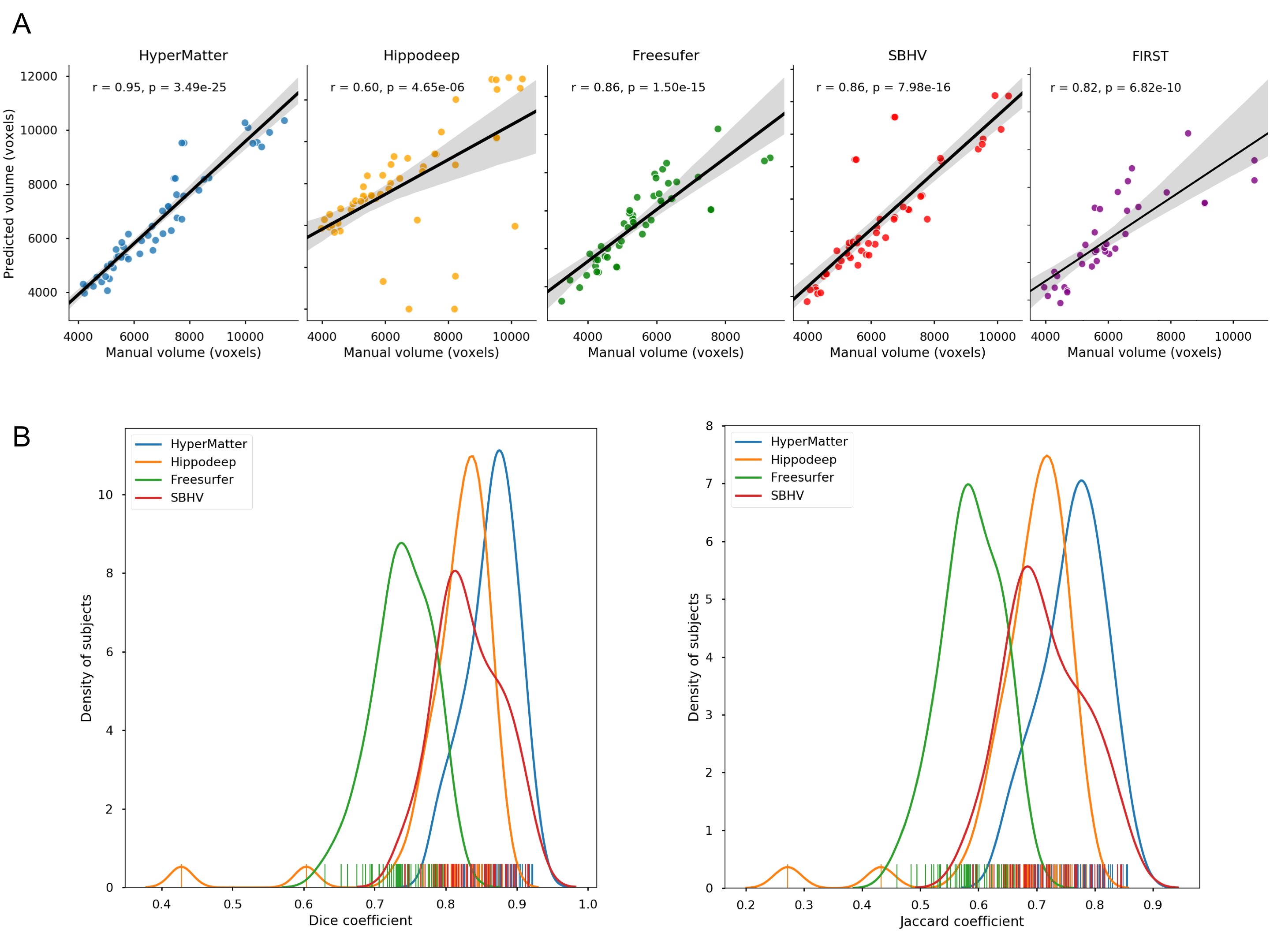
Validation
of hippocampal segmentation through volume correlations and Dice, Jaccard
similarity coefficients on our proposed model (HyperMatter - blue) and four
established techniques: Hippodeep - yellow, Freesurfer - green, SBHV - red and
FIRST - purple. The proposed model produced the best agreement to manual labels
among the five tested techniques in all three metrics. (A) Correlations between the manually
segmented volumes and volumes generated through model predictions. (B)
Distribution of Dice and Jaccard coefficients between ground truth manual
labels of the hippocampus and predicted segmentations.
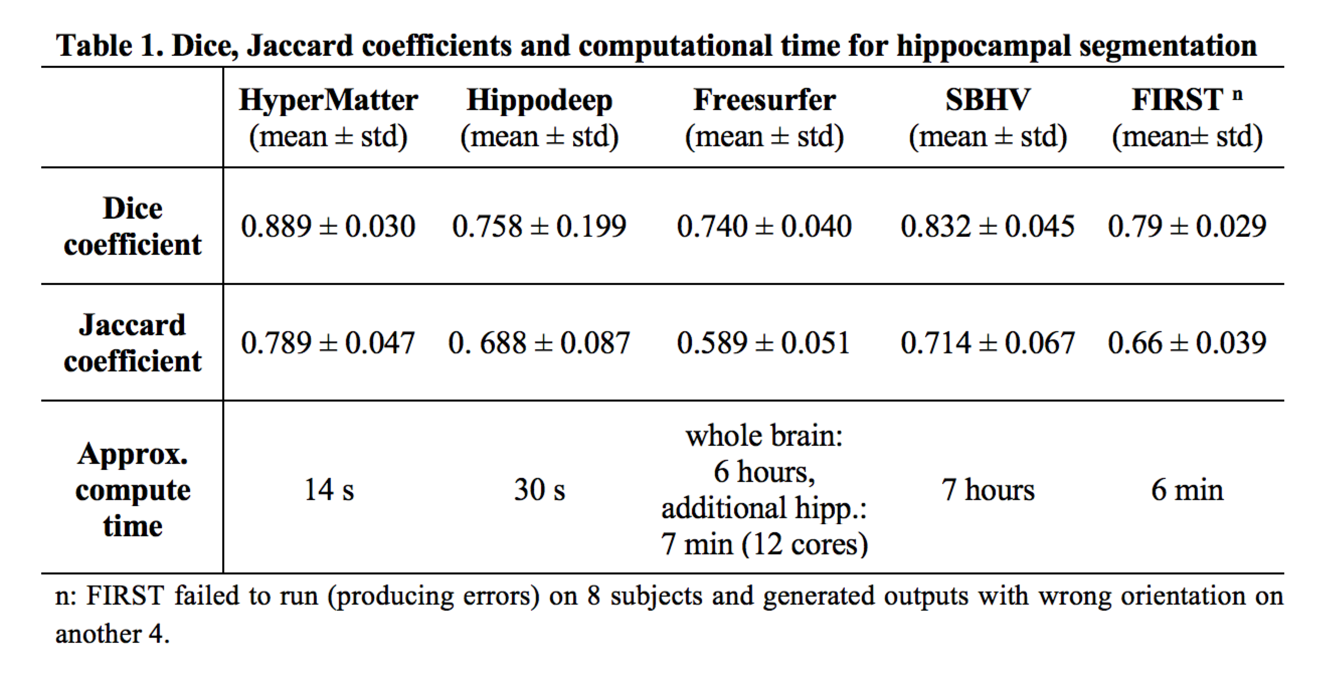
Table 1. Dice, Jaccard coefficients and computational time for the tested algorithms.
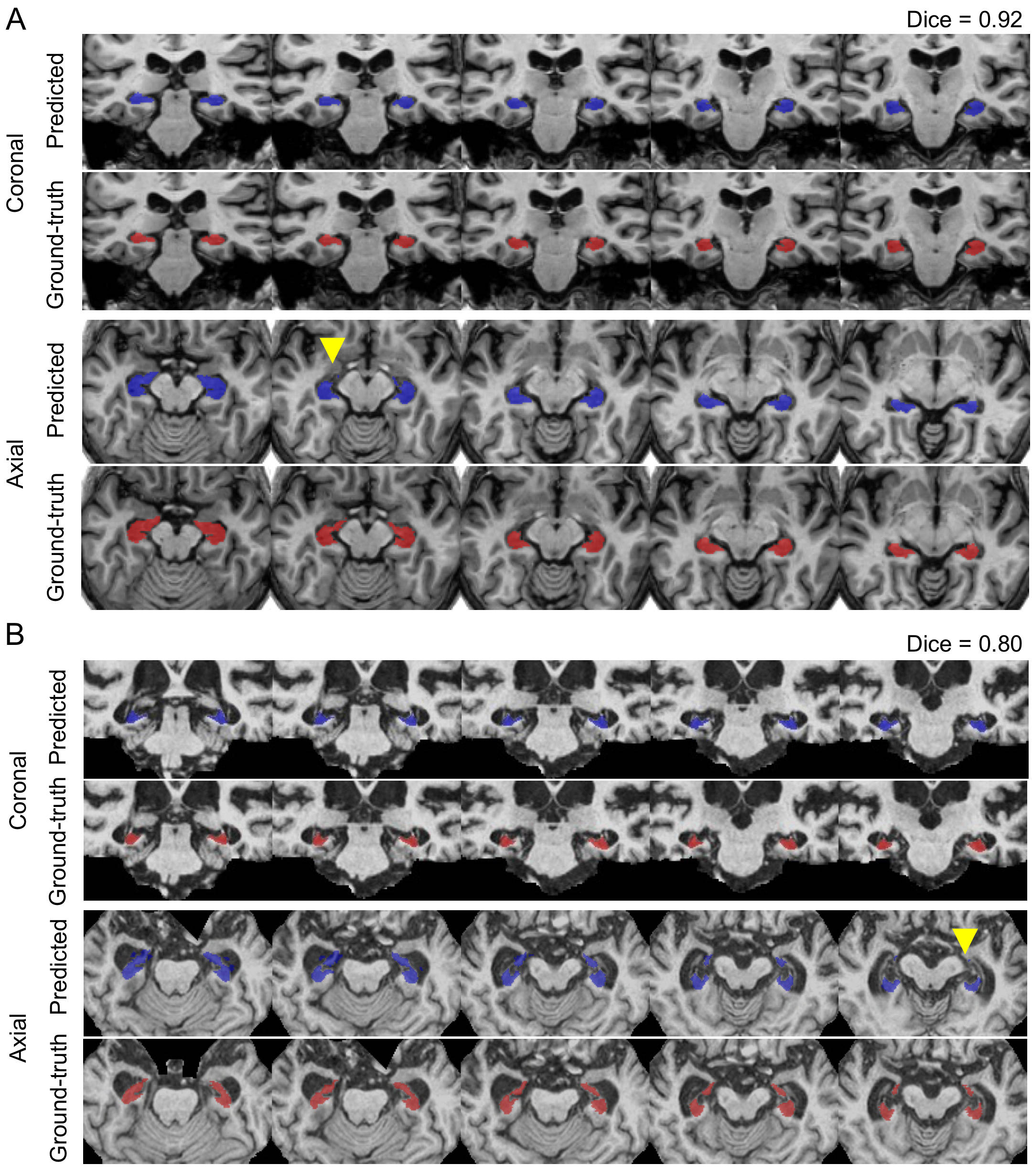
Hippocampal
segmentation cases with the highest (A) and lowest (B) dice coefficients from
the test set in coronal and axial views. Blue labels represent predicted
segmentations and red represent manual delineations. Yellow arrowheads
highlight areas of mis-segmentations.
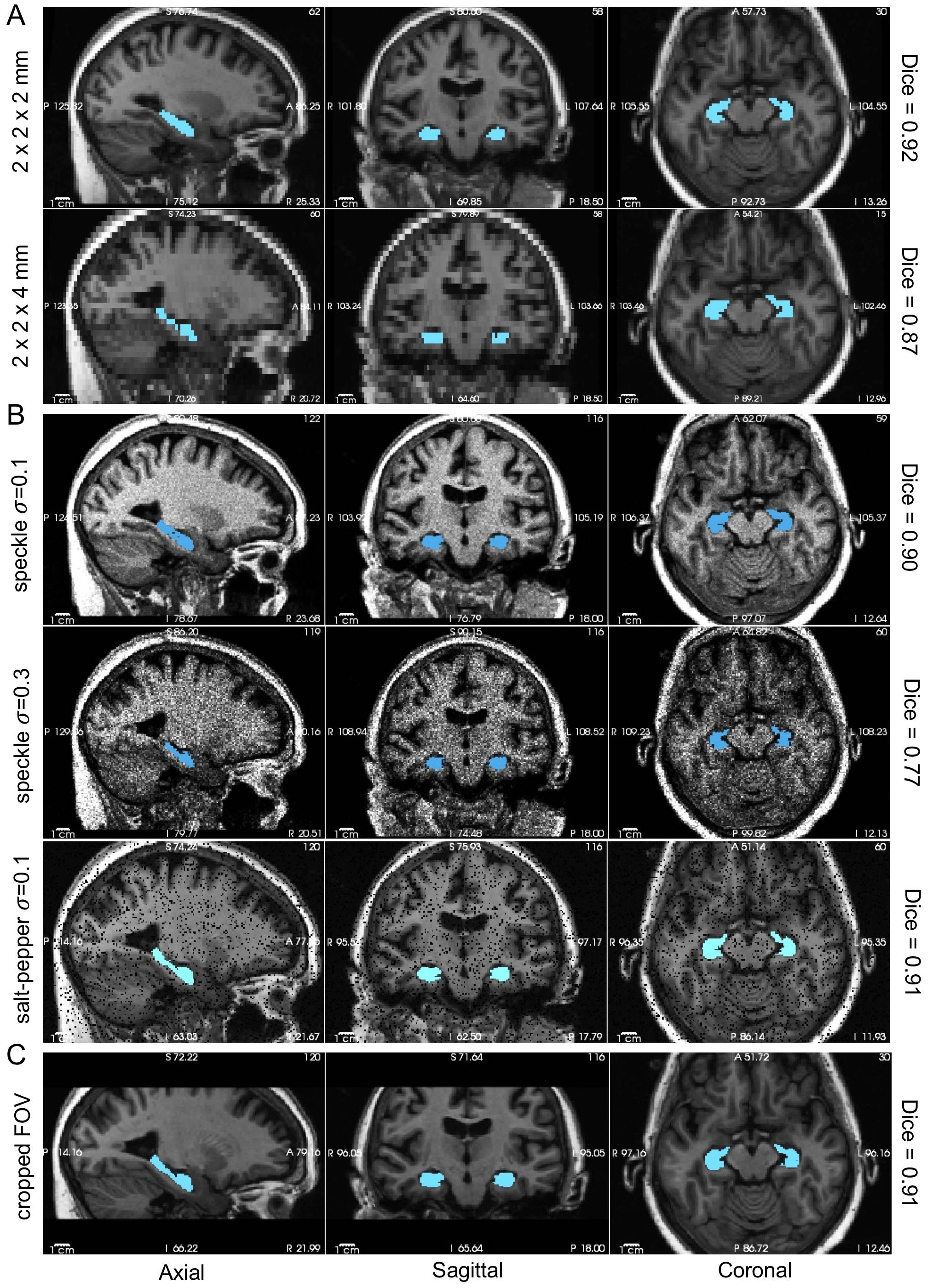
Adversarial
attacks on the hippocampal model to simulate clinical data with low resolution,
SNR and limited FOV, demonstrating the robustness of our model to such attacks. (A) Downsampling
of resolution. (B) Addition
of different amounts (sigmas) of speckle and salt-and-pepper noise to simulate
lower SNR. (C)
Cropping of FOV