0022
Automatic segmentation and follow-up of optic pathway gliomas using deep learning and based on conventional MRI1Sagol Brain Institute, Tel Aviv Sourasky Medical Center, Tel - Aviv, Israel, 2Sackler Faculty of Medicine, Tel Aviv University, Tel - Aviv, Israel, 3Sagol School of Neuroscience, Tel Aviv University, Tel - Aviv, Israel, 4The Iby and Aladar Fleischman Faculty of Engineering, Tel Aviv University, Tel - Aviv, Israel, 5Division of Radiology, Tel Aviv Sourasky Medical Center, Tel - Aviv, Israel, 6The Gilbert Israeli Neurofibromatosis Center, Tel - Aviv, Israel, 7Department of Pediatric Neurosurgery, Tel - Aviv, Israel
Synopsis
Optic pathway gliomas (OPG) are heterogeneous tumors with complex shape. The aim of this study was to implement a deep-learning approach for automatic segmentation and follow-up of patients with OPG based on conventional MRI. A total of 354 MRI scans from 53 patients where included. A neural-network with a U-net architecture was trained for segmentation of lesion area. The similarity coefficient score between segmentation results and ground truth was 0.812±0.159, with sensitivity=0.799±0.188, specificity=0.999±0.002 and correlation of r=0.987 (p<0.001) between lesion volumes. These results demonstrate the potential applicability of the proposed method for automatic radiological follow-up of patients with OPG.
Introduction
Optic pathway gliomas (OPG) account for 3-5% of all pediatric central nervous system tumors and represent the most common intrinsic optic nerve tumors and are the most common tumors of the central nervous system in patients with Neurofibromatosis-1 (NF1)(1). Although OPG are mostly histologically benign their growth may cause severe complications depending on location, size and characteristics, and their behavior can show an aggressive clinical course. MRI is the modality of choice for follow-up of OPG. The radiological criteria generally use linear measurements of the lesion area based on post contrast T1 weighted images (2,3), which is inaccurate, time-consuming, and may compromise disease follow-up. The aim of this study was to implement a deep learning approach based on conventional MRI for automatic segmentation and follow-up of patients with OPG.Methods
Patients and MRI Protocol: A total of 354 MRI scans obtained from 53 patients with OPG (22 males, age 6.9±5.3 years) were included. Patients were scanned longitudinally as part of their routine clinical assessment. Scans included T2 weighted images (T2WI) acquired before and T1 weighted images acquired after contrast agent injection (T1WI+C).
Image Analysis: Manual segmentation of the entire lesion area was performed using AnalyzeDirect software. The manual segmentations were used as ground truth for model training and final evaluation of the segmentation results. Further analysis was performed using Matlab 2018a and fast.ai platform (PyTorch environment) and included:
Dataset Preparation: Included realignment between the T1WI+C and T2WI and resizing of all images within the data set to a 256X256 pixel size, resulting in a total of 2498 slices. To increase the size of our dataset, data augmentation was performed and included rotation flipping and random lighting.
Network architecture: A U-Net based deep convolutional network was used with training and validation size of 1998 and 500 slices respectively and a batch size of 5. For the network training adaptive moment estimator (ADAM)(4) stochastic gradient based optimizer was used with learning rate = 0.05 and the maximum number of epochs = 8.
Evaluation of the segmentation results: The automatically generated lesion area was compared with the manually segmented lesion based on visual inspection, similarity between the components’ contours based on dice similarity coefficient (DSC) score, correlations and mean sensitivity, specificity and accuracy between the ground truth and the obtained lesion areas.
Results and Discussion
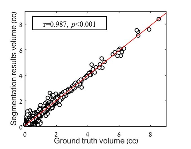
Segmentation results: In the majority of cases the proposed method accurately segmented OPG tumors based on conventional MRI. The average results for the entire lesion area were: DSC = 0.812±0.159, with sensitivity = 0.799±0.188, specificity = 0.999±0.002 accuracy = 0.997±0.003 and correlation of r=0.987 (p<0.001, Figure1) obtained between ground truth and the automated DL based segmentation results.
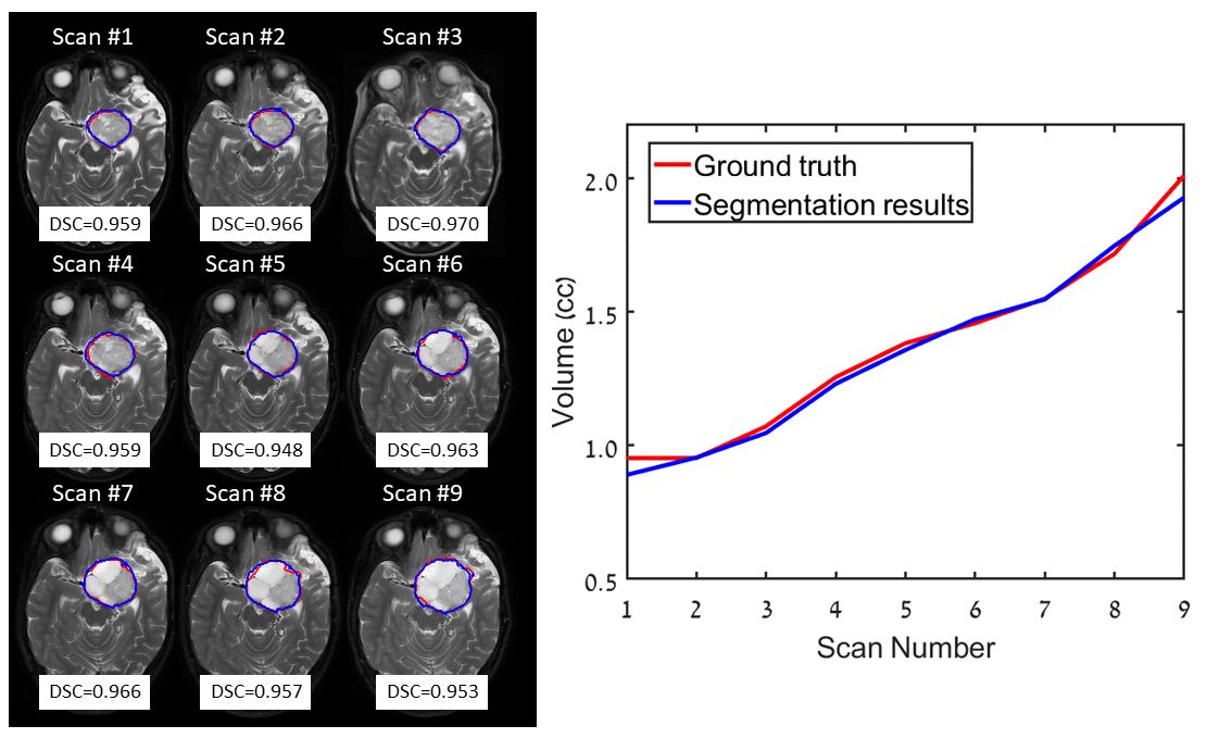
Longitudinal follow-up: Figure 2 shows longitudinal segmentation results obtained in a four year old female with OPG, scanned over five years (total of nine scans), demonstrating the high similarity obtained between ground truth (red) and automatic segmentation results (blue). This case demonstrates the importance of volumetric assessment in such patients. Only mild changes were detected between two sequential time points, (0-17%), which are difficult or impossible to identify based on visual assessment alone. However the longitudinal quantitative assessment revealed an ongoing pattern of increased lesion volume. The high similarity obtained in this case between the manual and the DL segmentation results (mean DSC=0.96±0.007) shows the potential applicability of this method to be integrated into routine clinical assessment of patients with ONG.
Conclusion
This study presents a DL based method for the automatic segmentation of OPG from conventional MR. The method resulted in accurate segmentation of this complex lesion area in the majority of cases, and enables automatic longitudinal assesment. An automatic tool for OPG volometric assesment can save much time and effort for clinicians and serve as an aid to support decision making in patients with OPG. We are currently performing optimization of the proposed method and sub segmentation of lesion area into its different components (enhancing, non-enhancing and cystic).Acknowledgements
No acknowledgement found.References
- Hudson S. Neurofibromatosis: historical perspective, classification and diagnostic criteria. The Neurofibromatoses A Pathogenetic and Clinical Overview 1994:1-22.
- Fried I, Tabori U, Tihan T, Reginald A, Bouffet E. Optic pathway gliomas: a review. CNS oncology 2013;2(2):143-159.
- Gnekow A. Recommendations of the Brain Tumor Subcommittee for the reporting of trials. Medical and pediatric oncology 1995;24(2):104-108.
- Kingma DP, Ba J. Adam: A method for stochastic optimization. arXiv preprint arXiv:14126980 2014.
Figures