0018
AUTOMATED BRAIN TISSUE SEGMENTATION USING DEEP LEARNING AND IMPERFECT LABELING1Radiology, University of Texas Southwestern Medical Center, Dallas, TX, United States
Synopsis
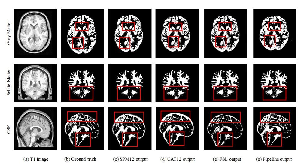
This work presents a deep learning pipeline to perform brain tissue segmentation on T1w Magnetic Resonance images (MRI). Two separate 3D-Dense-Unets were designed: GW-net to segment the gray matter (GM) and white matter (WM) and CSF-net to segment the cerebrospinal fluid (CSF). The network was trained on T1w MRI from 785 datasets in the iTAKL study with their corresponding SPM12 segmentations as ground truth and tested on 50 held-out subjects from the iTAKL study, 50 subjects from the AADHS study and 131 subjects from the Human Connectome project (HCP). Our pipeline showed improved segmentations when tested on simulated data with known ground truth as compared to the existing neuroimaging packages including SPM12, FSL and CAT12.
INTRODUCTION
Brain segmentation is an important image processing task and a key component in analyzing Magnetic Resonance (MR) images. Segmentation is an inherent part of voxel based morphometry [1], a widely used neuroimaging technique for analyzing MR images. Well-established segmentation tools such as FSL [2], SPM [1], Freesurfer [3] and others [4] have been developed [5]. These tools exhibit different strengths and weaknesses [6, 7] and brain researchers are left to choose knowing that different tools may introduce variable biases [8] and can negatively impact findings and weaken any drawn conclusions [5]. Here, we develop a deep learning pipeline which includes a dual network framework for automatic and accurate quantification of brain tissue segmentation.MATERIAL & METHODS
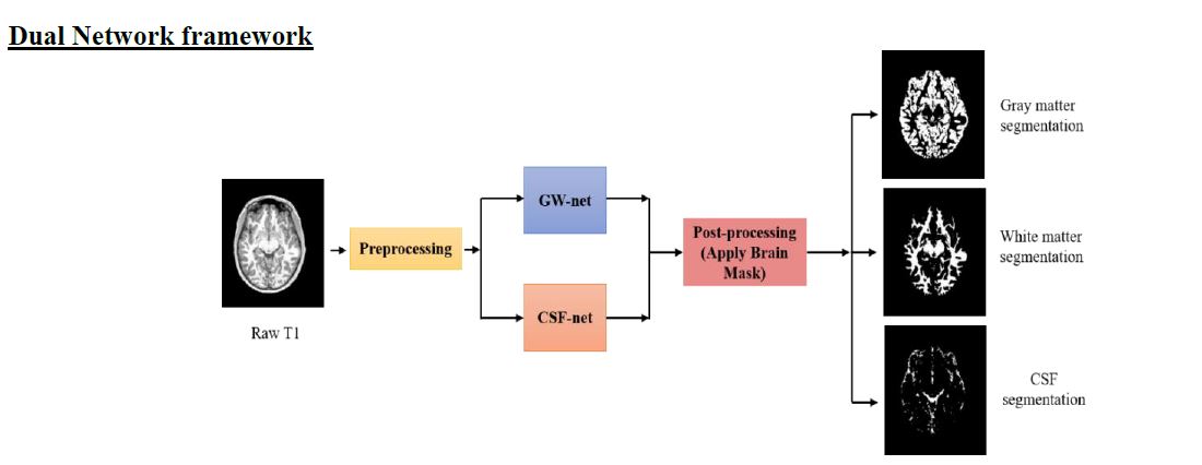
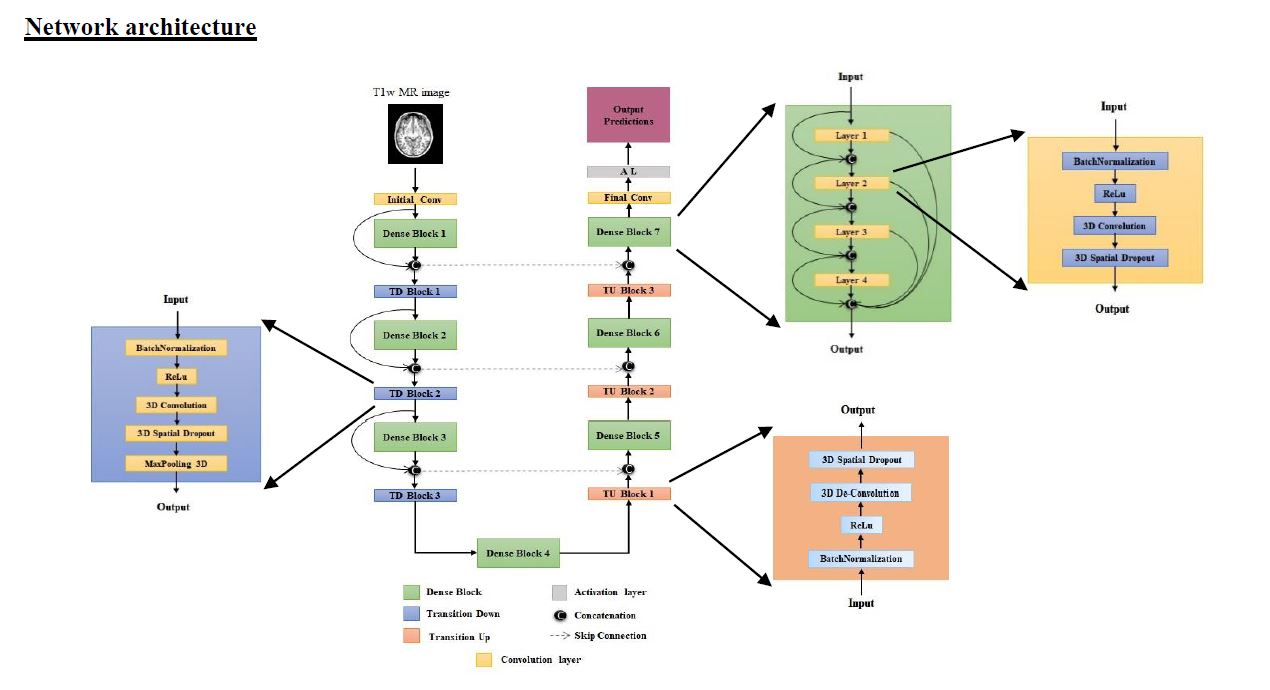
785 T1w MR images from male football players as part of the iTAKL[9] study of sub concussive impacts were used including 288 high school (14-18 years) and 497 youth (9-13 years) scans. Native space 3-class ground truth labels for all subjects were generated using SPM12 [1]. For the deep learning network, standard data preprocessing steps of the T1-weighted images were performed including: 1) N4BiasCorrection [10] to remove the RF inhomogeneity and 2) intensity normalization to 0-mean and unit variance. Two 3D-Dense-Unets were constructed to predict grey matter (GM), white matter (WM) and cerebrospinal fluid (CSF) segmentations. A 32x32x32 patch-based training and testing approach was implemented. 735 scans (268 high school and 467 youth) with their corresponding SPM12 tissue segmentations as ground truth were used for training the networks. Two networks were trained, 1) GM and WM segmentation and 2) CSF segmentation. The pipeline was tested on 50 held-out subjects from the iTAKL study [1], and 50 subjects from the AADHS study (a study of diabetes in African American adults). The network was additionally fine-tuned on 84 subjects from the HCP and tested on 131 held-out subjects from the HCP [11, 12]. For accurate quantification of the pipeline’s performance, it was then tested on simulated T1w MRI from BrainWeb [13, 14][15-19], with known ground truth. We developed 2 patch based 3D Dense-Unets to perform this task. A small pipeline that consists of 2 separate 3D-Dense-Unets to decompose the multi-class segmentation problem into binary segmentation problems was developed. Convolutional Neural Networks (CNNs) reduce the input resolution through multiple successive pooling layers and are suited well for applications where a single prediction per input image is expected. GW-net generates GM & WM segmentations, and CSF-net generates a CSF segmentation. A brain-mask was then applied using a 3D CNN trained for skull stripping to remove false positives outside the brain volume.RESULTS
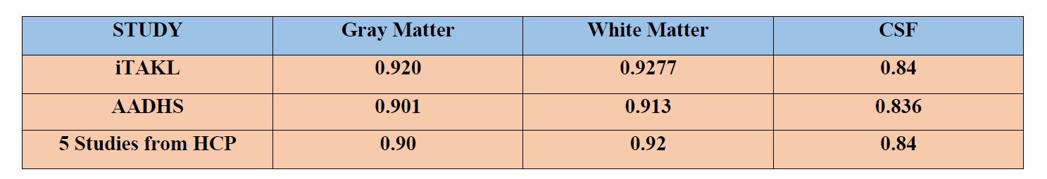
The pipeline performance on 50 subjects from the iTAKL study, 50 subjects from the AADHS study and 131 subjects from 5 studies from the HCP are tabulated below.
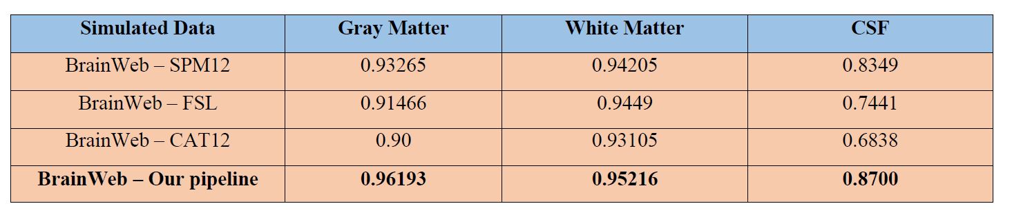
As the SPM12 segmentation maps were assumed to be perfect and used as ground truth for training the network, for accurate quantification of the pipeline’s performance, it was tested on simulated T1w MRIs from BrainWeb[13, 14][15-19], where the ground truth was known. Our pipeline segmented brain tissues with high accuracy outperforming SPM12, FSL and CAT12 in every tissue type with dice scores of 0.96193, 0.95216 and 0.84 for GM, WM and CSF respectively.
CONCLUSION
We demonstrate that our pipeline outperformed SPM12, FSL and CAT12 in brain tissue segmentation. In addition, the model does not require any initial skull-stripping which removes a potential source of error in data processing. The pipeline took approximately 5 minutes to segment the tissues whereas other packages took at least 10 minutes making it at least 2 times faster than these software packages.Acknowledgements
No acknowledgement found.References
1. Ashburner, J. and K.J. Friston, Voxel-based morphometry--the methods. Neuroimage, 2000. 11(6 Pt 1): p. 805-21. 2. Jenkinson, M., et al., Fsl. Neuroimage, 2012. 62(2): p. 782-90. 3. Dale, A.M., B. Fischl, and M.I. Sereno, Cortical surface-based analysis. I. Segmentation and surface reconstruction. Neuroimage, 1999. 9(2): p. 179-94. 4. Ledig, C., et al., Robust whole-brain segmentation: application to traumatic brain injury. Med Image Anal, 2015. 21(1): p. 40-58. 5. Rajchl, M., Pawlowski, Nick., Rueckert, Daniel., Matthews, Paul. M., Glocker, Ben, NeuroNet: Fast and Robust Reproduction of Multiple Brain Image Segmentation Pipelines. 1st Conference on Medical Imaging with Deep Learning (MIDL 2018), Amsterdam, The Netherlands., 2018. 6. Kazemi, K. and N. Noorizadeh, Quantitative Comparison of SPM, FSL, and Brainsuite for Brain MR Image Segmentation. J Biomed Phys Eng, 2014. 4(1): p. 13-26. 7. Heinen, R., et al., Robustness of Automated Methods for Brain Volume Measurements across Different MRI Field Strengths. PLoS One, 2016. 11(10): p. e0165719. 8. Tudorascu, D.L., et al., Reproducibility and Bias in Healthy Brain Segmentation: Comparison of Two Popular Neuroimaging Platforms. Front Neurosci, 2016. 10: p. 503. 9. Davenport, E.M., et al., Abnormal white matter integrity related to head impact exposure in a season of high school varsity football. J Neurotrauma, 2014. 31(19): p. 1617-24. 10. Tustison, N.J., et al., Large-scale evaluation of ANTs and FreeSurfer cortical thickness measurements. Neuroimage, 2014. 99: p. 166-79. 11. Mennes, M., et al., Making data sharing work: the FCP/INDI experience. Neuroimage, 2013. 82: p. 683-91. 12. Van Essen, D.C., et al., The WU-Minn Human Connectome Project: an overview. Neuroimage, 2013. 80: p. 62-79. 13. Aubert-Broche, B., A.C. Evans, and L. Collins, A new improved version of the realistic digital brain phantom. Neuroimage, 2006. 32(1): p. 138-45. 14. Aubert-Broche, B., et al., Twenty new digital brain phantoms for creation of validation image data bases. IEEE Trans Med Imaging, 2006. 25(11): p. 1410-6. 15. http://www.bic.mni.mcgill.ca/brainweb/ 16. C.A. Cocosco, V. Kollokian, R.K.-S. Kwan, A.C. Evans : "BrainWeb: Online Interface to a 3D MRI Simulated Brain Database" NeuroImage, vol.5, no.4, part 2/4, S425, 1997 -- Proceedings of 3-rd International Conference on Functional Mapping of the Human Brain, Copenhagen, May 1997. 17. R.K.-S. Kwan, A.C. Evans, G.B. Pike : "MRI simulation-based evaluation of image-processing and classification methods" IEEE Transactions on Medical Imaging. 18(11):1085-97, Nov 1999. 18. R.K.-S. Kwan, A.C. Evans, G.B. Pike : "An Extensible MRI Simulator for Post-Processing Evaluation" Visualization in Biomedical Computing (VBC'96). Lecture Notes in Computer Science, vol. 1131. Springer-Verlag, 1996. 135-140. 19. D.L. Collins, A.P. Zijdenbos, V. Kollokian, J.G. Sled, N.J. Kabani, C.J. Holmes, A.C. Evans : "Design and Construction of a Realistic Digital Brain Phantom" IEEE Transactions on Medical Imaging, vol.17, No.3, p.463--468, June 1998.Figures